. 2021 Jun 1;134(11):jcs255919.
doi: 10.1242/jcs.255919. Epub 2021 Jun 4.

Urokinase-type plasminogen activator-mediated crosstalk between N-cadherin and β-catenin promotes wound healing

Affiliations

Urokinase-type plasminogen activator-mediated crosstalk between N-cadherin and β-catenin promotes wound healing

Ariel Diaz et al. J Cell Sci. .

Abstract

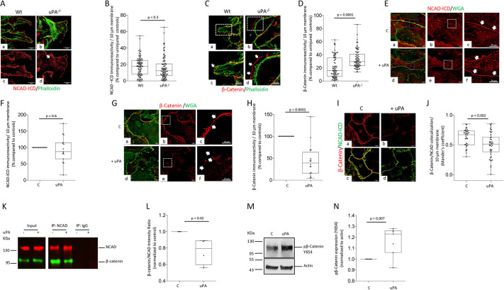
Urokinase-type plasminogen activator (uPA; encoded by Plau) is a serine proteinase that, in the central nervous system, induces astrocytic activation. β-Catenin is a protein that links the cytoplasmic tail of cadherins to the actin cytoskeleton, thus securing the formation of cadherin-mediated cell adhesion complexes. Disruption of cell-cell contacts leads to the detachment of β-catenin from cadherins, and β-catenin is then degraded by the proteasome following its phosphorylation by GSK3β. Here, we show that astrocytes release uPA following a scratch injury, and that this uPA promotes wound healing via a plasminogen-independent mechanism. We found that uPA induces the detachment of β-catenin from the cytoplasmic tail of N-cadherin (NCAD; also known as CDH2) by triggering its phosphorylation at Tyr654. Surprisingly, this is not followed by degradation of β-catenin because uPA also induces the phosphorylation of the low density lipoprotein receptor-related protein 6 (LRP6) at Ser1490, which then blocks the kinase activity of GSK3β. Our work indicates that the ensuing cytoplasmic accumulation of β-catenin is followed by its nuclear translocation and β-catenin-triggered transcription of the receptor for uPA (Plaur), which in turn is required for uPA to induce astrocytic wound healing.

Keywords: Low density lipoprotein receptor-related protein 6; Plasmin; Urokinase-type plasminogen activator; Wnt-β-catenin pathway; uPA; β-catenin.


Conflict of interest statement

Competing interests The authors declare no competing or financial interests.

Figures

Fig. 1.
Fig. 1.
uPA promotes astrocytic growth following a mechanical injury. (A) Representative live micrographs (10× objective magnification) of a monolayer of Wt astrocytes 0 and 24 h after a mechanical injury. White arrows in lower panel depict examples of zones of astrocytic growth in previously injured areas. (B) Mean concentration of uPA (ng/ml) in the culture medium of a monolayer of Wt astrocytes either left intact (control, C) or 24 h after a mechanical injury (I). n=7 (C) and n=6 (I). Unpaired two-tailed Student's t-test. (C) Representative live micrographs (10× objective magnification) of a monolayer of Wt astrocytes 0 and 24 h after a mechanical injury. Red lines depict the border of the wounded area immediately after the injury (upper panel) and the area of growth (lower panel) 24 h later. (D) Growth of a monolayer of Wt (n=8) and uPA−/− astrocytes (n=10) 24 h after a mechanical injury. Results are presented as percentage growth of the monolayer of uPA−/− astrocytes compared the growth of the Wt monolayer. Unpaired two-tailed Student's t-test. (E) Representative confocal micrographs viewed at 60× objective magnification of GFAP and Hoechst 33259 expression in the frontal cortex of Wt and uPA−/− mice 72 h after a mechanical injury. Scale bars: 50 μm. (F) GFAP-immunoreactive area per 4500 µm2 of cerebral cortex of Wt and uPA−/− mice (n=4) at 72 h after a mechanical injury. Each dot represents the average GFAP-positive area quantified in 5 micrographs per animal. Two-way ANOVA with Tukey's multiple comparisons test. (G) Growth of a monolayer of Wt and uPA−/− astrocytes 24 h after a mechanical injury and treatment with 5 nM of uPA [n=15 (Wt) and n=19 (uPA−/−)] or vehicle [control, n=15 (Wt) and n=19 (uPA−/−)]. Results are presented as percentage compared to growth of Wt astrocytes treated with vehicle (control). Two-way ANOVA with Tukey's multiple comparisons test. (H) Representative confocal micrographs viewed at 60× objective magnification of the injured border of two astrocytic monolayers stained with Hoechst 33259 (blue) and anti-KI67 antibodies (green) 24 h after a mechanical injury and treatment with vehicle (control; panels a,c) or 5 nM of uPA (panels b,d). White squares in panels a,b depict examples of KI67-positive astrocytes electronically magnified 10× in panels c,d. Scale bars: 100 μm (top panels), 20 μm (bottom panels). (I) Percentage of KI67-positive astrocytes in relation to the total number of cells in the injured border of astrocytic monolayers from four different cultures subjected to the experimental conditions described in H. Each dot represents the average value for each individual culture. n=221 (control) and 358 cells (uPA-treated). Unpaired two-tailed Student's t-test. (J) Growth of a monolayer of Wt astrocytes 24 h after a mechanical injury and treatment with vehicle (control; n=9), or 5 nM of uPA (n=11), or 100 nM of AraC (n=10), or a combination of uPA and AraC (n=12). Two-way ANOVA with Tukey's multiple comparisons test. (K) Astrocytic migration (µm/24 h) following a mechanical injury to a monolayer of Wt astrocytes in the presence of vehicle (control; n=14 cuts) or 5 nM of uPA (n=15 cuts). Each dot represents the average migration of 5 cells in the injured border. Unpaired two-tailed Student's t-test. (L) Astrocytic migration through a 5 μm pore insert following 24 h of treatment with 5 nM of uPA or a comparable volume of vehicle (control). Values are given as percentage of cells in the underside of the insert in relation to the number of cells in the upper side, normalized to values in vehicle (control)-treated cells. n=5 inserts assembled with astrocytes from three different cultures. Unpaired two-tailed Student's t-test. In all box plots, the box represents the 25–75th percentiles, and the median is indicated. The whiskers show the range.
Fig. 2.
Fig. 2.
uPA promotes astrocytic healing via a plasminogen-independent mechanism. (A) Growth of a monolayer of Wt and Plg−/− astrocytes 24 h after a wound injury and treatment with 5 nM of uPA [n=27 (Wt) and 27 (Plg−/−)] or vehicle [control; n=37 (Wt) and 39 (Plg−/−)]. Results are presented as percentage compared to growth of Wt astrocytes treated with vehicle (control). Unpaired two-tailed Student's t-test. (B) Growth of a monolayer of Wt astrocytes 24 h after a wound injury and treatment with vehicle (control, C, n=11) or 5 nM of uPA ATF (n=14). Unpaired two-tailed Student's t-test. (C) Growth of a monolayer of Wt astrocytes 24 h after a wound injury and treatment with vehicle (control, C, n=19) or 100 nM of plasmin (n=21). Unpaired two-tailed Student's t-test. (D) Growth of a monolayer of PAI-1−/− astrocytes (n=13) 24 h after a wound injury. Results are presented as percentage compared to growth of a monolayer of astrocytes prepared from Wt littermate controls (n=22) exposed to similar experimental conditions. Unpaired two-tailed Student's t-test. In all box plots, the box represents the 25–75th percentiles, and the median is indicated. The whiskers show the range.
Fig. 3.
Fig. 3.
uPA triggers the detachment of β-catenin from the intracellular domain of N-cadherin. (A) Representative confocal micrographs (viewed at 400×) from the wounded border of a monolayer of Wt (panels a,c) and uPA−/− astrocytes (panels b,d) stained with phalloidin (green) and antibodies against the intracellular domain of N-cadherin (NCAD-ICD; red) 24 h after a mechanical injury. Scale bars: 20 μm. (B) NCAD-ICD immunoreactivity per 10 μm of membrane of cells located in the wounded border of a monolayer of Wt (n=90) and uPA−/− (n=90) astrocytes 24 h after a mechanical injury. Results are presented as NCAD-ICD immunoreactivity per 10 μm of membrane compared to NCAD-ICD immunoreactivity in uninjured astrocytes. Unpaired two-tailed Student's t-test. (C) Representative confocal micrographs (viewed at 400×) from the wounded border of a monolayer of Wt (panels a,c) and uPA−/− astrocytes (panels b,d) stained with phalloidin (green) and anti-β-catenin antibodies (red) 24 h after a mechanical injury. Panels c,d correspond to a 2.5× electronic magnification of the area depicted with dashed squares in a,b. Arrows in c,d depict β-catenin-immunoreactive areas on the cell surface. Scale bars: 20 μm (top panels), 10 μm (bottom panels). (D) β-catenin immunoreactivity per 10 μm of membrane of cells along the wounded border of a monolayer of Wt (n=79) and uPA−/− (n=74) astrocytes 24 h after a mechanical injury. Results are presented in relation to β-catenin immunoreactivity in uninjured astrocytes. Unpaired two-tailed Student's t-test. (E–H) Representative confocal micrographs viewed at 400× (E,G) of monolayers of uninjured Wt astrocytes treated for 30 min with vehicle (control; C) or 5 nM of uPA, and then stained with WGA (green) and antibodies against either NCAD-ICD (red in E) or β-catenin (red in G). Panels c,f in E,G correspond to a 2.5× electronic magnification of the areas demarcated by the dashed squares in b,e. Arrows in c,f denote membrane staining for NCAD-ICD (E) or β-catenin (G). F,H correspond to NCAD-ICD (F) and β-catenin immunoreactivity (H) on the plasma membrane of uninjured Wt astrocytes treated with vehicle (control; n=15 cells from three different cultures) or 5 nM of uPA (n=15 cells from three different cultures). Unpaired two-tailed Student's t-test. Scale bars: 20 μm (E, a,b,d,e; G, a,b,d,e), 10 μm (E, c,f; G, c,f). (I) Representative confocal micrographs (viewed at 400×) of a monolayer of uninjured Wt astrocytes immunostained with antibodies against NCAD-ICD (green) and β-catenin (red) following 30 min of treatment with vehicle (control, C, panels a,c) or 5 nM of uPA (panels b,d). Scale bars: 20 μm. (J) β-catenin/NCAD colocalization per 10 μm of membrane (Mander's coefficient) in a monolayer of uninjured Wt astrocytes incubated for 30 min with 5 nM of uPA or vehicle (control, C). Unpaired two-tailed Student's t-test. (K,L) Extracts prepared from Wt astrocytes incubated during 30 min with 5 nM of uPA or vehicle (control) were immunoprecipitated with anti-NCAD and IgG antibodies and immunoblotted with antibodies against either β-catenin or NCAD. L depicts the ratio for β-catenin:NCAD intensity of the band in four observations per experimental group. Unpaired two-tailed Student's t-test. (M,N) Representative western blot analysis (M) and mean intensity of the band (N) of β-catenin phosphorylated at Tyr654 (pβ-catenin) in Wt astrocytes incubated for 15 min with 5 nM of uPA or vehicle (control). n=7. Unpaired two-tailed Student's t-test. In all box plots, the box represents the 25–75th percentiles, and the median is indicated. The whiskers show the range.
Fig. 4.
Fig. 4.
uPA inhibits the degradation of β-catenin. (A–F) Representative western blot analyses (A,C,E) and quantification of the intensity of the band (B,D,F) of total β-catenin (A–D), and active β-catenin (E,F) in Wt astrocytes incubated for 30 min (A,C), or for 0, 15 and 30 min (E) with vehicle (control, C) or 5 nM of either uPA (A,E) or its ATF (C). n=5 (B,D) and n=6 (F). Unpaired two-tailed Student's t-test was performed in B,D; one-way ANOVA with Dunnett's multiple comparisons test in F. (G) Mean growth of a monolayer of Wt astrocytes 24 h after a mechanical injury and treatment with vehicle (control; n=17), or 5 nM of uPA (n=25), or a combination of 5 nM of uPA and 1 μM of XAV-939 (n=24), or with XAV-939 alone (n=19). Results are presented as percentage of growth of wounded vehicle (control)-treated Wt astrocytes. Two-way ANOVA with Holm-Sidak's multiple comparisons test. In all box plots, the box represents the 25–75th percentiles, and the median is indicated. The whiskers show the range.
Fig. 5.
Fig. 5.
uPA induces LRP6 phosphorylation. (A–F) Representative western blot analyses (A,C,E) and the corresponding quantification of the mean intensity of the band (B,D,F) of LRP6 phosphorylated at Ser1490 (pLRP6) in Wt (A,C,E) and uPAR−/− astrocytes (C) incubated for 15 min with uPA or vehicle (control), alone or in the presence of 10 μM of the ERK 1/2 inhibitor SL327 (E,F). n=5 (B) and n=6 (D,F). Data are compared in each experiment to pLRP6 expression in Wt astrocytes treated with vehicle (control). Unpaired two-tailed Student's t-test. In all box plots, the box represents the 25–75th percentiles, and the median is indicated. The whiskers show the range.
Fig. 6.
Fig. 6.
uPA induces the nuclear translocation of β-catenin. (A,B) Representative western blot analyses (A) and quantification of the intensity of the band (B) of β-catenin phosphorylated at Ser675 in Wt astrocytes treated for 30 min with 5 nM of uPA or vehicle (control). n=5. Unpaired two-tailed Student's t-test. (C) Representative western blot analysis of β-catenin, GAPDH and histone-3 expression in cytosolic (Cyt) and nuclear (N) extracts prepared from Wt astrocytes treated for 30 min with 5 nM of uPA or vehicle (control). (D) β-catenin expression normalized to histone-3 expression in nuclear extracts prepared from Wt astrocytes exposed to the experimental conditions described in C. n=7. Unpaired two-tailed Student's t-test. (E) Panels a and d are representative confocal micrographs viewed at 60× objective magnification of β-catenin expression (green) in Wt astrocytes treated for 30 min with vehicle (control; panel a) or 5 nM of uPA (panel d). Panels b, c, e and f correspond to a 2.5× magnification of the area depicted by the dashed squares in a,d. Blue in b,e is Hoechst 33259. Arrows in c,f depict the absence (c) or presence (f) of β-catenin in the nucleus. Scale bars: 20 μm. (F) β-catenin-immunoreactive area in nuclei of Wt astrocytes treated for 30 min with vehicle (control; n=58 cells from three different cultures) or 5 nM of uPA (n=57 cells from three different cultures). Results are presented as percentage compared to vehicle (control)-treated cells. Mann–Whitney U test. In all box plots, the box represents the 25–75th percentiles, and the median is indicated. The whiskers show the range.
Fig. 7.
Fig. 7.
uPA induces β-catenin signaling. (A,D) Representative RT-PCR analyses of Myc (c-myc), Plaur and Actb mRNA expression in Wt astrocytes treated 24 h with 5 nM of uPA, or a comparable volume of vehicle (control), or with a combination of uPA and 1 μM of XAV-939, or with 1 µM of XAV-939 alone. (B,C,E,F) Mean Myc (B,E) and Plaur (C,F) mRNA expression, normalized to Actb mRNA expression and compared to the corresponding mRNA expression in vehicle (control)-treated Wt astrocytes. n=4 per experimental condition. Unpaired two-tailed Student's t-test (B,C) and two-way ANOVA with Holm–Sidak's multiple comparisons test (E,F). (G,H). Representative western blot analysis (G) and quantification of the intensity of the band (H) of uPAR expression in Wt astrocytes treated for 24 h with 5 nM of uPA or a comparable volume of vehicle (control). n=4. Unpaired two-tailed Student's t-test. (I) Growth of a monolayer of Wt and uPAR−/− astrocytes 24 h after a mechanical injury and treatment with 5 nM of uPA. Results are presented as percentage of growth compared to vehicle (control)-treated Wt astrocytes. n=32 (Wt treated with vehicle-control), n=43 [uPAR−/− treated with vehicle (control)], n=11 (uPA-treated Wt) and n=15 (uPA-treated uPAR−/−). Two-way ANOVA with Holm–Sidak's multiple comparisons test. In all box plots, the box represents the 25–75th percentiles, and the median is indicated. The whiskers show the range.
Fig. 8.
Fig. 8.
Proposed model of uPA-induced astrocytic healing. (A) The homophilic interaction between the extracellular domains of N-cadherin (NCAD-ECD), and the binding of β-catenin to the intracellular domain of NCAD (NCAD-ICD) mediate the formation of cell–cell contacts in a monolayer of uninjured astrocytes. (B–D) A mechanical injury that disrupts the interaction between the extracellular domains of NCAD (B) triggers the release of uPA. Binding of this uPA to its receptor (uPAR) induces the phosphorylation of β-catenin at Tyr654, thus prompting its detachment from NCAD-ICD (C). Simultaneously, uPA–uPAR binding-induced phosphorylation of the LRP6 intracellular domain (LRP6-ICD) at Ser1490, prompts the membrane recruitment of GSK3β, thus abrogating its ability to phosphorylate β-catenin. This not only allows β-catenin to escape proteasomal degradation, but also promotes its cytoplasmic accumulation and subsequent nuclear translocation, whereupon binding to TCF/Lef transcription factors triggers the expression of the uPA receptor (Plaur). Finally, by inducing the migration of cells from the injured border, the resultant increase in the abundance of uPAR on the plasma membrane promotes the healing of the wounded zone (D).

References

    1. Ahn, V. E., Chu, M. L.-H., Choi, H.-J., Tran, D., Abo, A. and Weis, W. I. (2011). Structural basis of Wnt signaling inhibition by Dickkopf binding to LRP5/6. Dev. Cell 21, 862-873. 10.1016/j.devcel.2011.09.003 - DOI - PMC - PubMed
    1. Asuthkar, S., Gondi, C. S., Nalla, A. K., Velpula, K. K., Gorantla, B. and Rao, J. S. (2012). Urokinase-type plasminogen activator receptor (uPAR)-mediated regulation of WNT/β-catenin signaling is enhanced in irradiated medulloblastoma cells. J. Biol. Chem. 287, 20576-20589. 10.1074/jbc.M112.348888 - DOI - PMC - PubMed
    1. Bafico, A., Liu, G., Yaniv, A., Gazit, A. and Aaronson, S. A. (2001). Novel mechanism of Wnt signalling inhibition mediated by Dickkopf-1 interaction with LRP6/Arrow. Nat. Cell Biol. 3, 683-686. 10.1038/35083081 - DOI - PubMed
    1. Bilic, J., Huang, Y.-L., Davidson, G., Zimmermann, T., Cruciat, C.-M., Bienz, M. and Niehrs, C. (2007). Wnt induces LRP6 signalosomes and promotes dishevelled-dependent LRP6 phosphorylation. Science 316, 1619-1622. 10.1126/science.1137065 - DOI - PubMed
    1. Blasi, F. and Carmeliet, P. (2002). uPAR: a versatile signalling orchestrator. Nat. Rev. Mol. Cell Biol. 3, 932-943. 10.1038/nrm977 - DOI - PubMed

Publication types

MeSH terms

Substances