. 2021 Aug;27(8):895-907.
doi: 10.1111/cns.13650. Epub 2021 Jun 4.

Chronic Toxoplasma gondii infection and sleep-wake alterations in mice

Affiliations

Chronic Toxoplasma gondii infection and sleep-wake alterations in mice

Damien Dupont et al. CNS Neurosci Ther. 2021 Aug.

Abstract

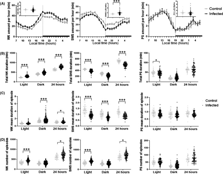
Aim: Toxoplasma gondii (Tg) is an intracellular parasite infecting more than a third of the human population. Yet, the impact of Tg infection on sleep, a highly sensitive index of brain functions, remains unknown. We designed an experimental mouse model of chronic Tg infection to assess the effects on sleep-wake states.

Methods: Mice were infected using cysts of the type II Prugniaud strain. We performed chronic sleep-wake recordings and monitoring as well as EEG power spectral density analysis in order to assess the quantitative and qualitative changes of sleep-wake states. Pharmacological approach was combined to evaluate the direct impact of the infection and inflammation caused by Tg.

Results: Infected mouse exhibited chronic sleep-wake alterations over months, characterized by a marked increase (>20%) in time spent awake and in cortical EEG θ power density of all sleep-wake states. Meanwhile, slow-wave sleep decreased significantly. These effects were alleviated by an anti-inflammatory treatment using corticosteroid dexamethasone.

Conclusion: We demonstrated for the first time the direct consequences of Tg infection on sleep-wake states. The persistently increased wakefulness and reduced sleep fit with the parasite's strategy to enhance dissemination through host predation and are of significance in understanding the neurodegenerative and neuropsychiatric disorders reported in infected patients.

Keywords: Toxoplasma; infection; neuropsychiatric disorders; sleep; wakefulness.


Conflict of interest statement

The authors declare no competing financial interests.

Figures

FIGURE 1
FIGURE 1
Chronology of experiments. Colored bands represent recording periods, colored arrows represent pharmacological administration of either dexamethasone or sulfamethoxazole‐trimethoprim. Time between recording periods consists of a washing period with no EEG nor EMG recordings
FIGURE 2
FIGURE 2
Comparison of spontaneous sleep‐wake parameters in non‐infected (up) and infected mice (low): Examples of polygraphic recordings and corresponding hypnograms showing the spontaneous sleep‐wake cycle
FIGURE 3
FIGURE 3
A, Sleep‐wake states amount per hour: Mean hourly values (± SEM in min) of the sleep‐wake states. The dark areas correspond to the dark period. The non‐background inserted dot plots correspond to the light/dark or dark/light (L/D or D/L) ratio of sleep‐wake amount before and after lights‐off. B, Total sleep‐wake states duration: Means ± SEM (in min) of the sleep‐wake stages for the 12 h light and dark and 24 h periods. C, Mean duration of episodes of each sleep‐wake state during the light, dark and 24 hours period (mean values ± SEM (in min)). D, Mean number of episode of each sleep‐wake state during the light, dark, and 24 hours period (mean values ± SEM). Other abbreviations: PS, Paradoxical Sleep; SWS, Slow‐Wave Sleep; WK, Waking. *P<0.05,***P<0.0001 two‐way ANOVA and unpaired Student's t test (A, inserted dot plots, two tails)
FIGURE 4
FIGURE 4
Influence of the time since infection on WK and SWS durations between infected (black) and control (uninfected) mice (gray), means ± SEM (in min) of the sleep‐wake stages for the 12 h dark and 24 h periods, showing an increase of WK with a decrease of SWS over time since infection. Time exacerbates the effects of Tg infection for WK total durations (dark: pinteraction Tg/time<0.0001, 24 hours: pinteraction Tg/time = 0.0013), as well as for SWS total duration during the dark period (pinteraction Tg/time<0.0001) and over 24 hours (pinteraction Tg/time = 0.011). Other abbreviations: Tg, Toxoplasma gondii; PS, Paradoxical Sleep; SWS, Slow‐Wave Sleep; WK, Waking. *P<0.05, ***P<0.0001 two‐way ANOVA followed by Holm‐Sidak multiple comparison post hoc tests
FIGURE 5
FIGURE 5
Mean spectral distribution of cortical EEG power density (X axis in Hz) in baseline sleep‐wake states in infected and control (uninfected) mice for each state (WK line 1, SWS line 2, PS line 3) and each period (24 hours column 1, light column 2, and dark column 3). The data were obtained by pooling consecutive 5 s epochs during 24 hours using the fast Fourier transform routine within the frequency range of 0.5‐20 Hz. For each curve, cortical EEG power band density (dot plot) is represented for infected (black) and control (gray) mice. Note that infected mice show an increase in theta band (6‐10 Hz) during all states and a decrease in slow activity in 1‐2 Hz range during all states. Other abbreviations: PS, Paradoxical Sleep; SWS, Slow‐Wave Sleep; WK, Waking. *P<0.05, ***P<0.0001 nonparametric Mann‐Whitney test (two tails)
FIGURE 6
FIGURE 6
A, Influence of the time since infection on WK durations between infected (black) and infected mice treated with DXM (purple), means ± SEM (in min) of the sleep‐wake stage for the 12 h light period, showing that increased WK due to infection is alleviated by DXM. This effect is impacted by time since infection. B, Relevant cortical EEG power band density (dot plot) is represented for infected (black) and infected+DXM (purple) mice during WK, vigilance state in which we observed major quantitative changes. Other abbreviations: pi, post‐infection; WK, Waking. *< 0.05,***< 0.0001 A/two‐way ANOVA followed by Holm‐Sidak multiple comparison post hoc tests and B/ nonparametric Mann‐Whitney test (two tails)
FIGURE 7
FIGURE 7
Graphic abstract illustrating the main findings of the present study and our interpretations. When mice are infected chronically by Toxoplasma gondii, an intracellular parasite, they exhibit a marked increase in wakefulness and activities, thus promoting encounters with cats, its natural predators. It is likely that by this strategy to modify its hosts’ sleep‐wake and behavioral states, Toxoplasma gondii would enhance its own dissemination, thus explaining why it infects a wide range of animals and about 1/3 of the world population

References

    1. Hill DE, Chirukandoth S, Dubey JP. Biology and epidemiology of Toxoplasma gondii in man and animals. Anim Health Res Rev. 2005;6(1):41‐61. 10.1079/ahr2005100 - DOI - PubMed
    1. Montoya JG, Liesenfeld O. Toxoplasmosis. Lancet. 2004;363(9425):1965‐1976. 10.1016/S0140-6736(04)16412-X - DOI - PubMed
    1. Berdoy M, Webster JP, Macdonald DW. Fatal attraction in rats infected with Toxoplasma gondii . Proc Biol Sci. 2000;267(1452):1591‐1594. 10.1098/rspb.2000.1182 - DOI - PMC - PubMed
    1. House PK, Vyas A, Sapolsky R. Predator cat odors activate sexual arousal pathways in brains of Toxoplasma gondii infected rats. PLoS One. 2011;6(8):e23277. 10.1371/journal.pone.0023277 - DOI - PMC - PubMed
    1. Ingram WM, Goodrich LM, Robey EA, Eisen MB. Mice infected with low‐virulence strains of Toxoplasma gondii lose their innate aversion to cat urine, even after extensive parasite clearance. PLoS One. 2013;8(9):e75246. 10.1371/journal.pone.0075246 - DOI - PMC - PubMed