. 2021 Aug 1;321(2):H259-H274.
doi: 10.1152/ajpheart.00174.2021. Epub 2021 Jun 4.

Cardiomyocyte-specific Txnip C247S mutation improves left ventricular functional reserve in streptozotocin-induced diabetic mice

Affiliations

Cardiomyocyte-specific Txnip C247S mutation improves left ventricular functional reserve in streptozotocin-induced diabetic mice

Nobuhiro Mukai et al. Am J Physiol Heart Circ Physiol. .

Abstract

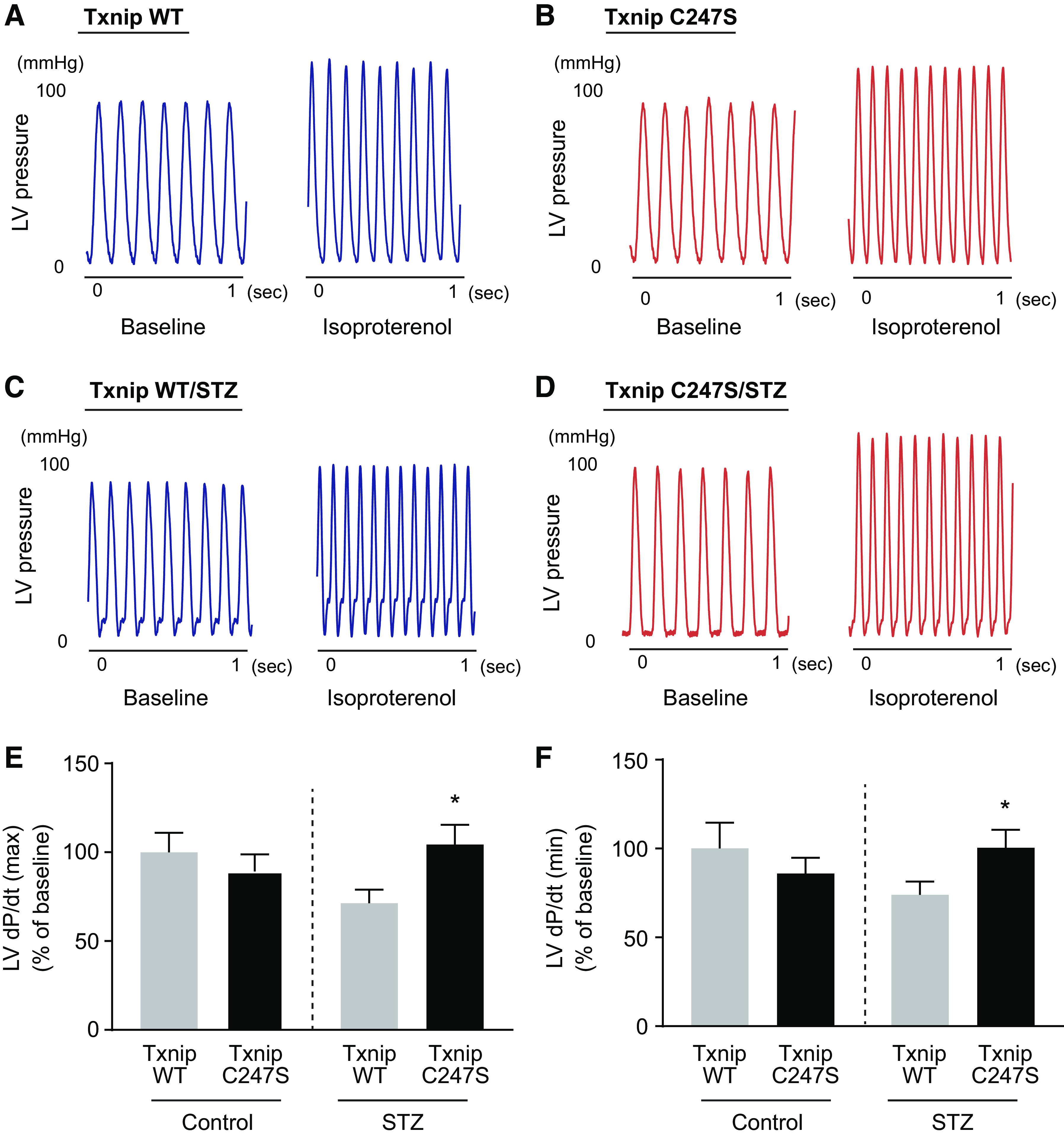
Underlying molecular mechanisms for the development of diabetic cardiomyopathy remain to be determined. Long-term exposure to hyperglycemia causes oxidative stress, which leads to cardiomyocyte dysfunction. Previous studies established the importance of thioredoxin-interacting protein (Txnip) in cellular redox homeostasis and glucose metabolism. Txnip is a highly glucose-responsive molecule that interacts with the catalytic center of reduced thioredoxin and inhibits the antioxidant function of thioredoxin. Here, we show that the molecular interaction between Txnip and thioredoxin plays a pivotal role in the regulation of redox balance in the diabetic myocardium. High glucose increased Txnip expression, decreased thioredoxin activities, and caused oxidative stress in cells. The Txnip-thioredoxin complex was detected in cells with overexpressing wild-type Txnip but not Txnip cysteine 247 to serine (C247S) mutant that disrupts the intermolecular disulfide bridge. Then, diabetes was induced in cardiomyocyte-specific Txnip C247S knock-in mice and their littermate control animals by injections of streptozotocin (STZ). Prolonged hyperglycemia upregulated myocardial Txnip expression in both genotypes. The absence of Txnip's inhibition of thioredoxin in Txnip C247S mutant hearts promoted mitochondrial antioxidative capacities in cardiomyocytes, thereby protecting the heart from oxidative damage by diabetes. Stress hemodynamic analysis uncovered that Txnip C247S knock-in hearts have a greater left ventricular contractile reserve than wild-type hearts under STZ-induced diabetic conditions. These results provide novel evidence that Txnip serves as a regulator of hyperglycemia-induced cardiomyocyte toxicities through direct inhibition of thioredoxin and identify the single cysteine residue in Txnip as a therapeutic target for diabetic injuries.NEW & NORTEWORTHY Thioredoxin-interacting protein (Txnip) has been of great interest as a molecular mechanism to mediate diabetic organ damage. Here, we provide novel evidence that a single mutation of Txnip confers a defense mechanism against myocardial oxidative stress in streptozotocin-induced diabetic mice. The results demonstrate the importance of Txnip as a cysteine-containing redox protein that regulates antioxidant thioredoxin via disulfide bond-switching mechanism and identify the cysteine in Txnip as a therapeutic target for diabetic cardiomyopathy.

Keywords: metabolism; reactive oxygen species; thioredoxin.


Figures

Figure 1.
Figure 1.
Txnip is a glucose-responsive protein that antagonizes antioxidative properties of thioredoxin through Txnip cysteine 247. A and B: HEK293T cells were transfected with Txnip wild-type (WT), or a single cysteine-to-serine mutation at Txnip Cys247 (C247S) fused with mCherry. To quench thiol-disulfide exchange reactions, cell lysates were treated with ice-cold 10% TCA. Then, free cysteines were alkylated with AMS to trap the complex between exogenous Txnip-mCherry fusion protein and endogenous thioredoxin protein. Lysates resolved by non-reducing SDS-PAGE were stained with anti-Txnip antibody JY2 or by an anti-thioredoxin-1 antibody. Control represents cellular lysate without transfection. Western analysis showed that Txnip C247S abolished the formation of the Txnip-thioredoxin complex. C: wild-type mouse embryonic fibroblasts (MEFs) were treated with 25 mM of d-glucose or d-mannitol for 3 h. Relative expression of Txnip mRNA was measured by real-time quantitative PCR and normalized to the level of GAPDH (N = 3). *P < 0.01 vs. baseline control and mannitol (25 mM). D: H9c2 cells were treated with indicated concentrations of d-glucose for 24 h. Western blot analysis was performed with the anti-Txnip antibody JY2. β-Actin and Coomassie blue staining serve as loading controls in the gel. E–H: Txnip, thioredoxin-1, and thioredoxin-2 expression levels were analyzed in wild-type MEFs treated with 25 mM of 2-deoxy-d-glucose (2DG) for 3 or 6 h (N = 3). Each value was normalized to the level of GAPDH. I and J: incubation with a high concentration of 2DG (25 mM) caused oxidative stress in wild-type MEFs (N = 3). Thioredoxin activity was measured with an insulin disulfide reduction assay. The positive 2',7'-dichlorodihydrofluorescein diacetate (H2DCFDA) staining in cultured cells was quantified as a marker of ROS using a microplate reader. *P < 0.01 vs. control. AMS, 4-acetamido-4′-maleimidylstilbene-2,2′-disulfonate; SH or HS, thiol; S-S, disulfide bond; MEFs, mouse embryonic fibroblasts; ROS, reactive oxygen species; Txnip, thioredoxin-interacting protein.
Figure 2.
Figure 2.
Thioredoxin activities in cardiomyocyte-specific Txnip C247S knock-in mouse model. Thioredoxin’s reducing activities were measured in cytosolic (A) and mitochondria (B)-enriched fractions from the heart and skeletal muscle tissues of mice at baseline. N = 3 each, P = N.S. C247S, single cysteine-to-serine mutation at Txnip cysteine 247; N.S., not significant; Txnip, thioredoxin-interacting protein; WT, wild type.
Figure 3.
Figure 3.
Diabetes is induced in cardiomyocyte-specific Txnip C247S knock-in mice. A: experimental protocol for induction of diabetes and Cre-recombination. Mice were received an intraperitoneal injection (i.p.) of streptozotocin (STZ; 50 mg/kg) for five consecutive days at the age of 5 wk old. Then, to generate temporally inducible cardiomyocyte-specific knock-in mutation of Txnip, diabetic αMHC-MerCreMer/Txnipflox/flox mice were injected with 0.5 mg of 4-hydroxytamoxifen per day for 10 days at the age of 7 wk old. Mice carrying the MerCreMer transgene and the Txnip flox allele, treated with vehicle lacking 4-hydroxytamoxifen, were used as Txnip wild-type (WT) control. B: upregulation of Txnip by hyperglycemia was confirmed by Western blot analysis in whole heart homogenates from Txnip WT and C247S knock-in mice. Total glucose transporter 1 (GLUT1) expression levels were not changed by hyperglycemia in Txnip WT and C247S knock-in hearts. C: eight weeks after induction of diabetes by STZ, blood glucose level was elevated in both genotypes. N = 15–18. *P < 0.01 vs. Control. D–F: cardiac hypertrophy was assessed by heart weight to tibial length ratio and histological myocyte cross-sectional area with periodic acid-Schiff (PAS) staining (N = 6). Heart weight to tibial length ratio was smaller in diabetic Txnip C247S hearts than in diabetic Txnip WT hearts (N = 15–18). *P < 0.05 vs. Txnip WT Control and WT/STZ. E and G: diabetes did not induce intestinal collagen depositions in the myocardium from both Txnip WT and C247S mice as assessed by Picrosirus red staining (N = 6). C247S, single cysteine-to-serine mutation at Txnip cysteine 247; Txnip, thioredoxin-interacting protein.
Figure 4.
Figure 4.
Cardiomyocyte-specific Txnip C247S hearts exhibit increased antioxidative properties against diabetic stress. A and B: thioredoxin activity was measured using an insulin disulfide reduction assay in the cytosolic and mitochondria-enriched fractions from heart homogenates (N = 4). *P < 0.05 vs. Txnip WT and C247S controls, †P < 0.01 vs. Txnip WT/STZ. C: the ratio of reduced glutathione (GSH) to oxidized glutathione (GSSG) was measured in the heart (N = 3). *P < 0.01 vs. Txnip WT and C247S controls, †P < 0.05 vs. Txnip WT/STZ. D: tissue levels of malondialdehyde, a product from lipid peroxidation, were measured as a biomarker of oxidative stress in heart homogenates (N = 4). *P < 0.01 vs. Txnip WT and C247S controls, and Txnip WT/STZ. E and F: frozen sections were stained with the green fluorescence dye H2DCFDA (4 µM) to assess the tissue levels of ROS. To confirm that H2DCFDA had specific staining, wild-type diabetic heart sections were preincubated with the O2· scavenger, tiron (10 mM). Then, H2DCFDA intensities in the myocardium were quantified and normalized over the background fluorescence (N = 3). G: redox-regulated gene expression of heme oxygenase-1 (HO-1) was measured by real-time quantitative PCR and normalized to the level of β-actin (N = 6–9). *P < 0.01 vs. Txnip WT and C247S controls, and †P < 0.05 vs. Txnip WT/STZ. C247S, single cysteine-to-serine mutation at Txnip cysteine 247; H2DCFDA, 2',7'-dichlorodihydrofluorescein diacetate; ROS, reactive oxygen species; STZ, streptozotocin; Txnip, thioredoxin-interacting protein.
Figure 5.
Figure 5.
Cardiomyocyte-specific Txnip C247S mutation prevents cardiomyocyte damage against diabetic stress. A–C: TUNEL-positive cardiomyocytes were determined by triple staining with TUNEL (green), anti-sarcomeric α actin antibody (red), and DAPI (blue). Active caspase-3 was detected with anti-cleaved caspase3 antibody (green) in paraffin-embedded heart tissues. N = 6. *P < 0.01 vs. Txnip WT and C247S controls, and †P < 0.05 vs. Txnip WT/STZ. D and E: vessel area within the left ventricular myocardium was quantified by immunostaining with Griffonia Simplicifolia isolectin B4 (red) and DAPI (blue). N = 6. P = N.S. C247S, single cysteine-to-serine mutation at Txnip cysteine 247; N.S., not significant; STZ, streptozotocin; Txnip, thioredoxin-interacting protein; WT, wild type.
Figure 6.
Figure 6.
A–H: stress hemodynamic analysis uncovers the evidence of masked mechanical cardiac dysfunction by hyperglycemia. Hearts were perfused in a Langendorff mode. Heart rate and left ventricular (LV) pressure were recorded during and after isoproterenol (iso, 0.05 µM) infusion. β-Adrenergic challenge increased heart rate after the injection in all hearts, but changes in LV pressure in response to isoproterenol were less in streptozotocin (STZ)-induced diabetic wild-type (WT) hearts than in non-diabetic WT hearts. N = 7 each for control and N = 8 each for STZ hearts. *P < 0.05 and †P < 0.01 vs. STZ by two-way ANOVA. C247S, single cysteine-to-serine mutation at Txnip cysteine 247; STZ, streptozotocin; Txnip, thioredoxin-interacting protein; WT, wild type.
Figure 7.
Figure 7.
Txnip C247S knock-in hearts have a greater inotropic reserve under diabetic conditions. A–D: representative tracing of left ventricular (LV) pressure in a nondiabetic control mouse heart and a diabetic heart from Txnip wild-type (WT) or C247S knock-in animal with a Langendorff perfusion system. E and F: the acute increases in left ventricular dP/dtmax and dP/dtmin from baseline to 2 min after isoproterenol (iso, 0.05 µM) infusion were significantly less in streptozotocin (STZ)-induced diabetic wild-type (WT) hearts than in nondiabetic WT hearts or in Txnip C247S hearts with or without diabetes. N = 7 each for control and N = 8 each for STZ hearts. *P < 0.05 vs. Txnip WT STZ. C247S, single cysteine-to-serine mutation at Txnip cysteine 247; STZ, streptozotocin; Txnip, thioredoxin-interacting protein; WT, wild type.
Figure 8.
Figure 8.
Schematic of the proposed mechanisms by which Txnip C247S achieves cardioprotection in diabetes. Hyperglycemia-induced Txnip decreases thioredoxin’s antioxidant activities and inhibits glucose utilization in cardiomyocytes. These molecular processes promote the development of diabetic cardiomyopathy. Txnip C247S mutant is incapable of binding thioredoxin but retains the ability to bind glucose transporter (GLUT). Unlike previous knockout models, our knock-in study specifically demonstrates a thioredoxin-dependent function of Txnip in the diabetic heart. C247S, single cysteine-to-serine mutation at Txnip cysteine 247; ROS, reactive oxygen species; Txnip, thioredoxin-interacting protein.

Comment in

Similar articles

Cited by

References

    1. Services USDoHaH. Estimates of diabetes and its burden in the United States. Centers for Disease Control and Prevention National Diabetes Statistics Report 2020: 1–30, 2020. CS314227-A. https://www.cdc.gov/diabetes/data/statistics-report/index.html.
    1. Schofield J, Ho J, Soran H. Cardiovascular risk in type 1 diabetes mellitus. Diabetes Ther 10: 773–789, 2019. doi:10.1007/s13300-019-0612-8. - DOI - PMC - PubMed
    1. Brownlee M. The pathobiology of diabetic complications: a unifying mechanism. Diabetes 54: 1615–1625, 2005. doi:10.2337/diabetes.54.6.1615. - DOI - PubMed
    1. Giacco F, Brownlee M. Oxidative stress and diabetic complications. Circ Res 107: 1058–1070, 2010. doi:10.1161/CIRCRESAHA.110.223545. - DOI - PMC - PubMed
    1. Yoshioka J. Thioredoxin superfamily and its effects on cardiac physiology and pathology. Compr Physiol 5: 513–530, 2015. doi:10.1002/cphy.c140042. - DOI - PubMed

Publication types

MeSH terms

LinkOut - more resources