Comparative Study
. 2021 Jun 1;62(7):6.
doi: 10.1167/iovs.62.7.6.

Expression of the SARS-CoV-2 Receptor ACE2 in Human Retina and Diabetes-Implications for Retinopathy

Affiliations
Comparative Study

Expression of the SARS-CoV-2 Receptor ACE2 in Human Retina and Diabetes-Implications for Retinopathy

Lingli Zhou et al. Invest Ophthalmol Vis Sci. .

Abstract

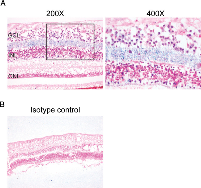
Purpose: To investigate the expression of angiotensin-converting enzyme 2 (ACE2), the receptor for SARS-CoV-2 in human retina.

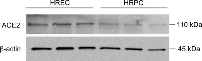
Methods: Human post-mortem eyes from 13 non-diabetic control cases and 11 diabetic retinopathy cases were analyzed for the expression of ACE2. To compare the vascular ACE2 expression between different organs that involve in diabetes, the expression of ACE2 was investigated in renal specimens from nondiabetic and diabetic nephropathy patients. Expression of TMPRSS2, a cell-surface protease that facilitates SARS-CoV-2 entry, was also investigated in human nondiabetic retinas. Primary human retinal endothelial cells (HRECs) and primary human retinal pericytes (HRPCs) were further used to confirm the vascular ACE2 expression in human retina.

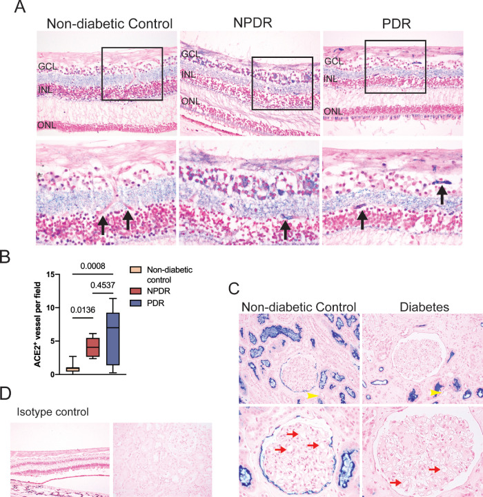
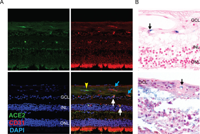
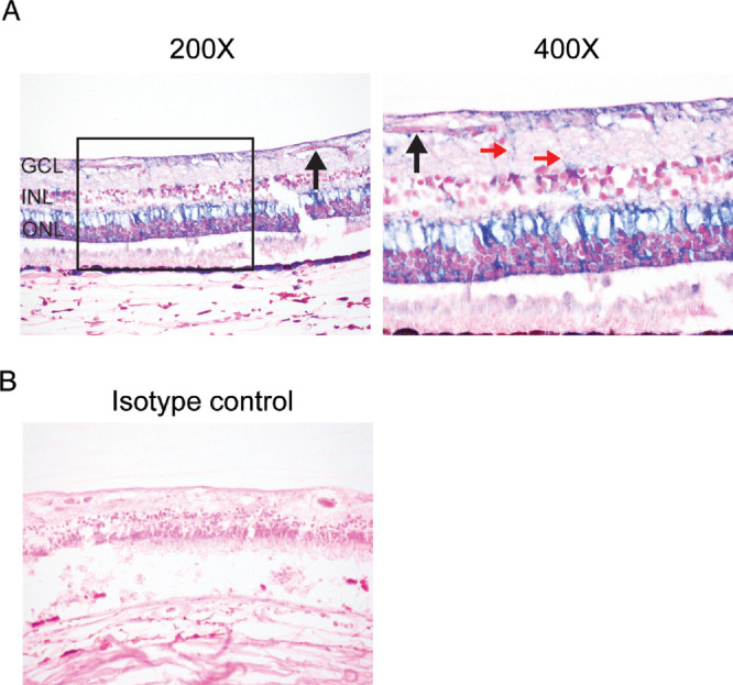
Results: We found that ACE2 was expressed in multiple nonvascular neuroretinal cells, including the retinal ganglion cell layer, inner plexiform layer, inner nuclear layer, and photoreceptor outer segments in both nondiabetic and diabetic retinopathy specimens. Strikingly, we observed significantly more ACE2 positive vessels in the diabetic retinopathy specimens. By contrast, in another end-stage organ affected by diabetes, the kidney, ACE2 in nondiabetic and diabetic nephropathy showed apical expression of ACE2 tubular epithelial cells, but no endothelial expression in glomerular or peritubular capillaries. Western blot analysis of protein lysates from HRECs and HRPCs confirmed expression of ACE2. TMPRSS2 expression was present in multiple retinal neuronal cells, vascular and perivascular cells, and Müller glia.

Conclusions: Together, these results indicate that retina expresses ACE2 and TMPRSS2. Moreover, there are increased vascular ACE2 expression in diabetic retinopathy retinas.


Conflict of interest statement

Disclosure: L. Zhou, None; Z. Xu, None; J. Guerra, None; A.Z. Rosenberg, None; P. Fenaroli, None; C.G. Eberhart, None; E.J. Duh, None

Figures

Figure 1.
Figure 1.
ACE2 expression in human retina. (A) Characteristic ACE2 expression in retina specimens from nondiabetic retina. ACE2 expression was found in some cells in the retinal ganglion cell layer, inner plexiform layer, and inner nuclear layer. (B) Isotype IgG control was negative for staining.
Figure 2.
Figure 2.
Increased vascular ACE2 expression in diabetic retinopathy retina. (A) Characteristic ACE2 expression in retina specimens from nondiabetic retina, NPDR, and PDR. Black arrows denote retinal capillaries, with significantly increased staining in diabetes. (B) ACE2 positive vessel staining was quantitated in retinas from nondiabetic retina, NPDR, and PDR. (C) Characteristic ACE2 expression in renal specimens from nondiabetic and diabetic nephropathy. ACE2 staining was absent in peritubular capillaries (yellow arrowhead) and glomerular capillary loop (red arrow). (D) Isotype IgG control was negative for staining.
Figure 3.
Figure 3.
ACE2 expression in vessels (A) ACE2 is colocalized with some CD31-positive endothelial cells in retina. White arrows show ACE2 positive capillaries in cross section. Blue arrows mark longitudinal cuts through a capillary with endothelial cells positive for both CD31 and ACE2; ACE2 expression in perivascular cells (yellow arrowhead); (B) Upper panel: ACE2 expression in a small capillary endothelial cell (arrow), with a larger caliber vessel above lined by ACE2-negative endothelial cells. Lower panel: ACE2 expression in a small capillary endothelial cell (arrow) in a hyalinized vessel.
Figure 4.
Figure 4.
ACE2 expression in primary human retina endothelial cells and primary human pericytes. Western blot analysis demonstrated ACE2 protein expression in both primary human retina endothelial cells and primary human pericytes.
Figure 5.
Figure 5.
TMPRSS2 expression in human retina. (A) Characteristic TMPRSS2 expression in retina specimens from nondiabetic retina. TMPRSS2 expression was found in some cells in the nerve fiber layer, retinal ganglion cell layer, inner nuclear layer, outer plexiform layer, and outer nuclear layer. TMPRSS2 is expressed in vascular/perivascular cells (black arrow) and radial processes morphologically consistent with Müller glia (red arrows). (B) Isotype IgG control was negative for staining.

Similar articles

Cited by

References

    1. Hoffmann M, Kleine-Weber H, Schroeder S, et al. .. SARS-CoV-2 cell entry depends on ACE2 and TMPRSS2 and is blocked by a clinically proven protease inhibitor. Cell. 2020; 181: 271–280.e278. - PMC - PubMed
    1. Walls AC, Park YJ, Tortorici MA, Wall A, McGuire AT, Veesler D.. Structure, function, and antigenicity of the SARS-CoV-2 spike glycoprotein. Cell. 2020; 181: 281–292.e286. - PMC - PubMed
    1. Gheblawi M, Wang K, Viveiros A, et al. .. Angiotensin-converting enzyme 2: SARS-CoV-2 receptor and regulator of the renin-angiotensin system: celebrating the 20th anniversary of the discovery of ACE2. Circ Res. 2020; 126: 1456–1474. - PMC - PubMed
    1. Patel VB, Zhong JC, Grant MB, Oudit GY.. Role of the ACE2/angiotensin 1-7 axis of the renin-angiotensin system in heart failure. Circ Res. 2016; 118: 1313–1326. - PMC - PubMed
    1. Miller AG, Tan G, Binger KJ, et al. .. Candesartan attenuates diabetic retinal vascular pathology by restoring glyoxalase-I function. Diabetes. 2010; 59: 3208–3215. - PMC - PubMed

Publication types

MeSH terms