. 2021 Jul 20;120(14):2828-2837.
doi: 10.1016/j.bpj.2021.05.026. Epub 2021 Jun 2.

Neuropilin-1 assists SARS-CoV-2 infection by stimulating the separation of Spike protein S1 and S2

Affiliations

Neuropilin-1 assists SARS-CoV-2 infection by stimulating the separation of Spike protein S1 and S2

Zhen-Lu Li et al. Biophys J. .

Abstract

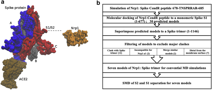
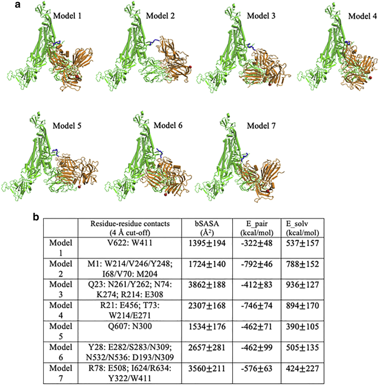
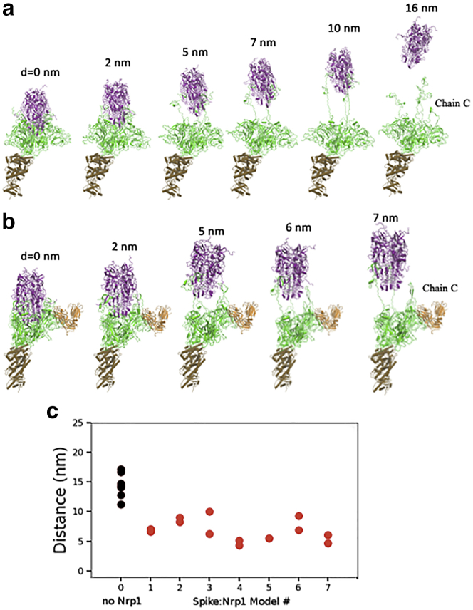
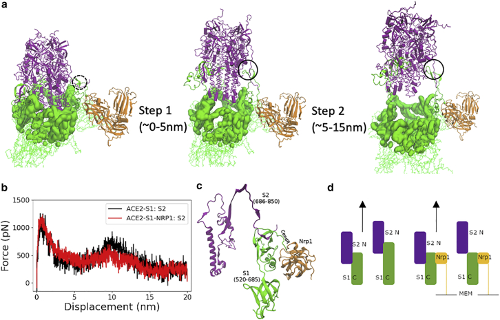
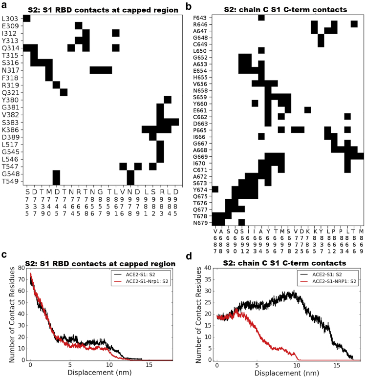
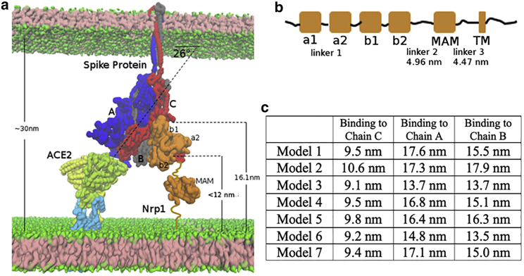
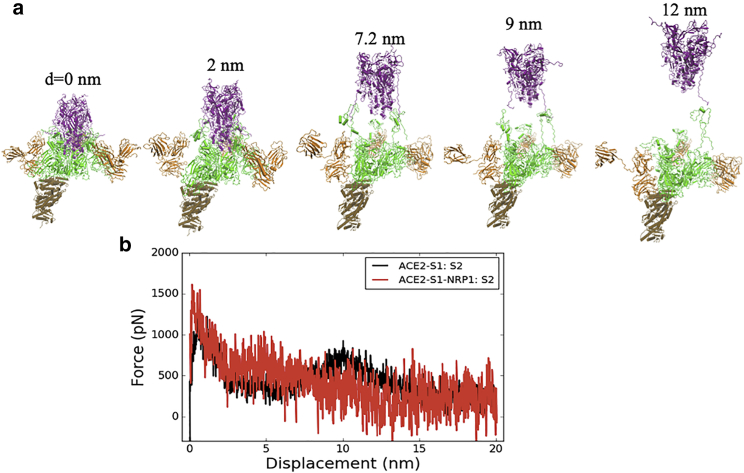
The cell surface receptor Neuropilin-1 (Nrp1) was recently identified as a host factor for severe acute respiratory syndrome coronavirus 2 (SARS-CoV-2) entry. The Spike protein of SARS-CoV-2 is cleaved into two segments, the S1 (residues (res.) 1-685) and the S2 (res. 686-1273) domains by furin protease. Nrp1 predominantly binds to the C-terminal RRAR amino acid motif (res. 682-685) of the S1 domain. In this study, we firstly modeled the association of an Nrp1 protein (consisting of domains a2-b1-b2) with the Spike protein. Next, we studied the separation of S2 from the S1 domain, with and without Nrp1 bound, by utilizing molecular dynamics pulling simulations. During the separation, Nrp1 stabilizes the S1 C-terminal region (res. 640-685) and thereby assists the detachment of S2 N-terminal region (res. 686-700). Without Nrp1 bound, S1 tends to become stretched, whereas the bound Nrp1 stimulates an earlier separation of S2 from the S1 domain. The liberated S2 domain is known to mediate the fusion of virus and host membranes; thus, Nrp1 likely increases virus infectivity by facilitating the S1 and S2 separation. We further analyzed the possible topological structure of the SARS-CoV-2 Spike protein when bound with Nrp1 and angiotensin-converting enzyme 2 (ACE2). Understanding of such an Nrp1-assisted viral infection opens the gate for the generation of protein-protein inhibitors, such as antibodies, which could attenuate the infection mechanism and protect certain cells in a future Nrp1-ACE2 targeted combination therapy.


Figures

Figure 1
Figure 1
Overview of a Spike protein trimer with ACE2 and Nrp1 and the workflow of the study. (a) ACE2 binds to Spike trimer RBD (chain A). Nrp1 associates with Spike trimer at the S1-S2 cleavage site (chain C). (b) Flow chart of the computational modeling and simulation approach utilized in this study. To see this figure in color, go online.
Figure 2
Figure 2
Binding modes for seven predicted models. (a) Models, shown for Spike trimer (chain-C): Nrp1 (a2-b1-b2) binding. The TNSPRRAR motif is marked in blue. The last residue of the Nrp1 a2-b1-b2 domain segment is marked in red. (b) Noticeable residue-residue contacts (<0.4 nm) between Spike protein and Nrp1 in different models (except for the RRAR binding region). Together with additional residue-residue contacts (<0.6 nm), the proteins are seen to interact via domains a2, b1, and b2 on Nrp1 and a limited number of regions N1–N4, RBDN, RBDC, C1–C3, the very C-terminus of S1 and beginning of S2 (see Fig. S3 for details). Listed in Table b are the buried solvent-accessible surface area (SASA), pair-interaction energy, and changes of solvation energy for the different models. The interaction energies (based upon pairwise vdW and electrostatic forces) are calculated between Nrp1 (domains a2-b1-b2) with a full Spike trimer (chains A, B, and C). The models shown are listed in order of their population near their cluster centers (most to least). Standard errors were calculated over different frames of the simulation trajectories (saved at every 100 ps). To see this figure in color, go online.
Figure 3
Figure 3
Separation process of the ACE2-S1 and S2 domain of chain C with and without Nrp1, here shown (a) in absence of Nrp1 (first of seven simulations shown) and (b) for ACE2-S1-Nrp1:S2 model 7, bottom. (S1 is in green, S2 in purple, and Nrp1 in orange.) The displacement of the S2 domain c.o.m relative to initial position is given as distance d. The time interval in the pulling simulations (1 nm/ns) between the structures shown is 2, 5, 7, 10, and 16 ns without Nrp1 bound and 2, 5, 6, and 7 ns with Nrp1 bound, respectively. (c) Plot of the distance between the S1 and the S2 c.o.ms of chain C at the time of separation (when nearest distance S1:S2 distance becomes >5 Å). Two simulations were run and reported for each model except for model 5, in which Nrp1:S1 binding is easily disconnected during the pulling of the S2 domain. To see this figure in color, go online.
Figure 4
Figure 4
Two-step separation between S2 and S1. (a) Two steps of S2 exit from the S1 domain: S1 regions that cap S2 domain (purple) are highlighted in a surface representation in green. (b) Pulling force versus displacement of S2 domain (with and without Nrp1). Results shown were averaged over duplicate simulations of the seven models without and with Nrp1. (c) S2-S1 contacts at the cleavage site at the start of the simulation (here model 7 at 0 ns). (d) Shows a schematic picture of the “holding effect” of Nrp1—essentially, Nrp1 binding stabilizes the region it associates with and does not allow the stretching of this region. (S2 is in violet, S1 in green, and Nrp1's primary binding domain b1 in yellow.)
Figure 5
Figure 5
Contacts between S2 and S1 at capped region and at the S1-S2 cleavage region. The cap region comprises S1 res. 303–549 (mostly RBD region) and S2 res. 735–768/856–985. The S1-S2 cleavage region consists of S1 res. 640–685 and S2 res. 686–700. (a) Contact map between S1 RBD and S2 subunit. (b) Contact map between S1 C-terminal region and S2. (c) Number of residues of S2 (chains A, B, and C) that interact with S1 RBD (chains A, B, and C) at a different position of S2 displacement with and without Nrp1 bound. (d) Number of residues of S2 (chains A, B, and C) that interact with S1 C-terminal region (res. 640–685 only of chain C), again with and without Nrp1 bound. Note that the number of contacts is counted to all three S protein units and temporarily increases as the S1-S2 cleavage region becomes distorted. To see this figure in color, go online.
Figure 6
Figure 6
Topological constraints of Nrp1 on binding with Spike protein trimer, bound to an ACE2 dimer. (a) ACE2 (yellow and lime, which also go into and through the membrane) and B0AT1 (light blue) 2:2 dimer complex at the bottom left. Shown is the Spike protein trimer (chains A, B, and C in blue, gray, and red, respectively) in the middle. Shown is Nrp1 (orange) at the bottom right of the figure. (b) Linear domain arrangement in the sequence of the Nrp1 receptor. (c) Estimated distance of C-terminus of the Nrp1 b2 domain to the membrane bilayer lipid headgroups in the different models with Nrp1 bound to chains A, B, and C. To see this figure in color, go online.
Figure 7
Figure 7
Separation process of S2 and S1 with three Nrp1s, i.e., one binding to each unit of the Spike protein trimer. (a) Snapshots and the displacement of the S2 domain relative to initial position are shown. (b) Pulling force versus displacement of the S2 domain with three Nrp1 bound. Data are shown as averaged over two simulations for model 1 (red), that is the separation of two times three S1:S2 contacts, whereas the ACE2 data are taken from Fig. 4b. To see this figure in color, go online.

Update of

Similar articles

Cited by

References

    1. Yan R., Zhang Y., Zhou Q. Structural basis for the recognition of SARS-CoV-2 by full-length human ACE2. Science. 2020;367:1444–1448. - PMC - PubMed
    1. Hoffmann M., Kleine-Weber H., Pöhlmann S. SARS-CoV-2 cell entry depends on ACE2 and TMPRSS2 and is blocked by a clinically proven protease inhibitor. Cell. 2020;181:271–280.e8. - PMC - PubMed
    1. Shang J., Wan Y., Li F. Cell entry mechanisms of SARS-CoV-2. Proc. Natl. Acad. Sci. USA. 2020;117:11727–11734. - PMC - PubMed
    1. Vankadari N. Structure of furin protease binding to SARS-CoV-2 spike glycoprotein and implications for potential targets and virulence. J. Phys. Chem. Lett. 2020;11:6655–6663. - PubMed
    1. Hoffmann M., Kleine-Weber H., Pöhlmann S. A multibasic cleavage site in the spike protein of SARS-CoV-2 is essential for infection of human lung cells. Mol. Cell. 2020;78:779–784.e5. - PMC - PubMed