. 2021 Aug:275:120842.
doi: 10.1016/j.biomaterials.2021.120842. Epub 2021 May 1.

Solubilized ubiquinol for preserving corneal function

Affiliations

Solubilized ubiquinol for preserving corneal function

Youssef W Naguib et al. Biomaterials. 2021 Aug.

Abstract

Defective cellular metabolism, impaired mitochondrial function, and increased cell death are major problems that adversely affect donor tissues during hypothermic preservation prior to transplantation. These problems are thought to arise from accumulated reactive oxygen species (ROS) inside cells. Oxidative stress acting on the cells of organs and tissues preserved in hypothermic conditions before surgery, as is the case for cornea transplantation, is thought to be a major reason behind cell death prior to surgery and decreased graft survival after transplantation. We have recently discovered that ubiquinol - the reduced and active form of coenzyme Q10 and a powerful antioxidant - significantly enhances mitochondrial function and reduces apoptosis in human donor corneal endothelial cells. However, ubiquinol is highly lipophilic, underscoring the need for an aqueous-based formulation of this molecule. Herein, we report a highly dispersible and stable formulation comprising a complex of ubiquinol and gamma cyclodextrin (γ-CD) for use in aqueous-phase ophthalmic products. Docking studies showed that γ-CD has the strongest binding affinity with ubiquinol compared to α- or β-CD. Complexed ubiquinol showed significantly higher stability compared to free ubiquinol in different aqueous ophthalmic products including Optisol-GS® corneal storage medium, balanced salt solution for intraocular irrigation, and topical Refresh® artificial tear eye drops. Greater ROS scavenging activity was noted in a cell model with high basal metabolism and ROS generation (A549) and in HCEC-B4G12 human corneal endothelial cells after treatment with ubiquinol/γ-CD compared to free ubiquinol. Furthermore, complexed ubiquinol was more effective at lowering ROS, and at far lower concentrations, compared to free ubiquinol. Complexed ubiquinol inhibited lipid peroxidation and protected HCEC-B4G12 cells against erastin-induced ferroptosis. No evidence of cellular toxicity was detected in HCEC-B4G12 cells after treatment with complexed ubiquinol. Using a vertical diffusion system, a topically applied inclusion complex of γ-CD and a lipophilic dye (coumarin-6) demonstrated transcorneal penetrance in porcine corneas and the capacity for the γ-CD vehicle to deliver drug to the corneal endothelium. Using the same model, topically applied ubiquinol/γ-CD complex penetrated the entire thickness of human donor corneas with markedly greater ubiquinol retention in the endothelium compared to free ubiquinol. Lastly, the penetrance of ubiquinol/γ-CD complex was assayed using human donor corneas preserved for 7 days in Optisol-GS® per standard industry practices, and demonstrated higher amounts of ubiquinol retained in the corneal endothelium compared to free ubiquinol. In summary, ubiquinol complexed with γ-CD is a highly stable composition that can be incorporated into a variety of aqueous-phase products for ophthalmic use including donor corneal storage media and topical eye drops to scavenge ROS and protect corneal endothelial cells against oxidative damage.

Keywords: Coenzyme Q10; Corneal endothelium; Cyclodextrin; Eye banking; Ferroptosis; Inclusion complex; Keratoplasty; Molecular docking; Reactive oxygen species; Ubiquinol.


Conflict of interest statement

Conflicts of Interest: No relevant financial interests to disclose.

Declaration of interests

The authors declare that they have no known competing financial interests or personal relationships that could have appeared to influence the work reported in this paper.

Figures

Figure 1:
Figure 1:
Molecular interactions between ubiquinol and α-, β- and γ-cyclodextrins. (A) 3D structures of α-, β-, and γ-cyclodextrins as soft surfaces, docked pose of ubiquinol in the cavity of α-, β-, and γ-cyclodextrins, interactions between ubiquinol and α-, β-, and γ-cyclodextrins with the H-bond distances, and representative hydrophobic interactions between ubiquinol and α-, β-, and γ-cyclodextrins with distances. Dashed green lines represent conventional H-bonds, dashed blue lines represent carbon-hydrogen bonds, and dashed magenta lines represent hydrophobic interactions. BIOVIA Discovery Studio Visualizer was used to generate H-bond distances, and PyMOL was used to generate the distances of hydrophobic interactions. (B) 3D structure of ubiquinol. (C) Predicted binding energy of ubiquinol to each of the three types of cyclodextrins used. (D) Phase solubility diagram of ubiquinol in different concentrations of γ-CD. (E) Photo: (on left): dispersion of 50 mg of ubiquinol/γ-CD complex (equivalent to 3.125 mg ubiquinol) added to 10 ml of water and shaken for 2 hours; (on right): dispersion of 5 mg ubiquinol alone added to 10 ml of water and shaken for 2 hours. (F) DSC thermograms and (G) XRD patterns of ubiquinol, γ-CD, ubiquinol/γ-CD physical mixture, and ubiquinol/γ-CD complex. (H) SEM images of ubiquinol, γ-CD, and ubiquinol/γ-CD complex. Top panel: X150, middle panel: X600 and bottom panel: X1200.
Figure 2:
Figure 2:
Stability of ubiquinol alone versus ubiquinol/γ-CD complex in (A) Optisol-GS® and (B) BSS. Stability is measured with regard to ubiquinol (the reduced form), ubiquinone (the oxidized form), and total coenzyme Q10. Values represent mean ± SD, * p < 0.05, ** p < 0.01, n = 3.
Figure 3:
Figure 3:
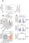
(A) Flow cytometric histograms of A549 cells. (B) and (C), bar graph figures representing the values obtained from the statistical analysis (geometric means) of the DHE fluorescence signals from histograms (values are means ± SD, **** p < 0.0001, *** p < 0.001, ** p < 0.01, and * p < 0.05). All groups represented by B and C were processed simultaneously, and they were split into two Figures to emphasize the specific message that each set conveys, and to improve clarity. (D) Amount of total CoQ10, ubiquinol and ubiquinone taken up into A549 cells after incubation of either free ubiquinol or ubiquinol/γ-CD complex with the cells for 1 or 3 hours at 37° C. Values represent mean ± SD, **p < 0.01, n = 4-6).
Figure 4:
Figure 4:
(A) Flow cytometric analysis following DHE-based ROS assay in HCEC-B4G12 cells. (B) DHE fluorescence signal values (means ± SD, **** p < 0.0001, *** p < 0.001, and ** p < 0.01) following the ROS assay in HCEC-B4G12 cells measured by flow cytometry (geometric means). Unless otherwise mentioned, all group were treated with antimycin A (AM; Fig. A and B). (C) MTS-based cytotoxicity assay of HCEC-B4G12 cells following 24 h and 72 h of incubation with different concentrations of ubiquinol, ubiquinol/γ-CD complex, and γ-CD (values are means ± SD). (D) Schematic showing the proposed role of ubiquinol to inhibit lipid peroxidation and the subsequent ferroptosis (Cys: cysteine, Glu: glutamate, GSH: reduced glutathione, GSSG: oxidized glutathione). (E) Flow cytometric analysis following C11-Bodipy 581/591-based lipid peroxidation assay in HCEC-B4G12 cells. (F) Bodipy 581/591 fluorescence signal values (means ± SD, **** p < 0.0001, *** p < 0.001, and ** p < 0.01) following lipid peroxidation assay in HCEC-B4G12 cells measured by flow cytometry (median). Unless otherwise mentioned, all group were treated with 10μM erastin (Fig. E and F). (G) MTS cytotoxicity assay of HCEC-B4G12 cells following erastin treatment (values are means ± SD, **** p < 0.0001). (H) Light microscopic images of cells treated with different treatment groups. (I) LDH-based cytotoxicity assay of HCEC-B4G12 cells (values are means ± SD, **** p < 0.0001).
Figure 5:
Figure 5:
(A) Coumarin-6/γ-CD complex (molar ratio of 1:10) shows much higher corneal penetrance compared to free coumarin-6 when applied to the corneal epithelium. Complexed coumarin-6 was able to diffuse uniformly across and through the anterior cornea from the epithelial side (top panel), and it was able to penetrate the entire cornea and reach the endothelial side (bottom panel) while free coumarin-6 could not. (B) Cumulative amount (ng) versus time of total coenzyme Q10 (ubiquinol and ubiquinone) detected in fluid on the endothelial side after permeation across human donor corneas fixed in a Navicyte vertical diffusion system after either ubiquinol/γ-CD complex or free ubiquinol (300 μM, n = 3/group) was added to the epithelial side of the diffusion apparatus. (C) Amount of total coenzyme Q10 (ng) retained in the endothelial cell Descemet membrane complex (EDM) of donor corneas isolated after permeation of either complexed or free ubiquinol (n = 4/group). (D) Amount of total coenzyme Q10 (ng) retained in the EDM of human donor corneas stored for 1 week (4° C) in Optisol-GS® supplemented with either complexed or free ubiquinol (10 μM, n = 4/group) in standard corneal storage containers (shown in the inset). Data represent average ± SD, p < 0.05 following two tailed Student’s T-test). (E) Pairwise comparison of complexed versus free ubiquinol uptake in the EDM of human donor corneas stored in Optisol-GS® supplemented storage media depicted in (D). For each donor pair, the right cornea was treated with complexed ubiquinol and the left cornea was treated with free ubiquinol.

References

    1. Awad EM, Khan SY, Sokolikova B, Brunner PM, Olcaydu D, Wojta J et al. Cold induces reactive oxygen species production and activation of the NF-kappa B response in endothelial cells and inflammation in vivo. Journal of thrombosis and haemostasis : JTH. 2013;11(9):1716–26. doi:10.11n/jth.12357. - DOI - PubMed
    1. Eltzschig HK, Eckle T. Ischemia and reperfusion-from mechanism to translation. Nature medicine. 2011;17(11):1391–401. doi:10.1038/nm.2507. - DOI - PMC - PubMed
    1. Yellon DM, Hausenloy DJ. Myocardial reperfusion injury. The New England journal of medicine. 2007;357(11): 1121–35. doi:10.1056/NEJMra071667. - DOI - PubMed
    1. Philipponnet C, Aniort J, Garrouste C, Kemeny JL, Heng AE. Ischemia reperfusion injury in kidney transplantation: A case report. Medicine. 2018;97(52):e13650. doi:10.1097/MD.0000000000013650. - DOI - PMC - PubMed
    1. Park SW, Kim M, Brown KM, D’Agati VD, Lee HT. Paneth cell-derived interleukin-17A causes multiorgan dysfunction after hepatic ischemia and reperfusion injury. Hepatology. 2011;53(5):1662–75. doi:10.1002/hep.24253. - DOI - PMC - PubMed

Publication types