

Differences in the clinicopathological features of pancreatic head carcinoma in dorsal and ventral pancreas: A single institution retrospective review
- PMID: 34087876
- PMCID: PMC8183761
- DOI: 10.1097/MD.0000000000026167
Differences in the clinicopathological features of pancreatic head carcinoma in dorsal and ventral pancreas: A single institution retrospective review
Abstract
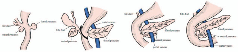
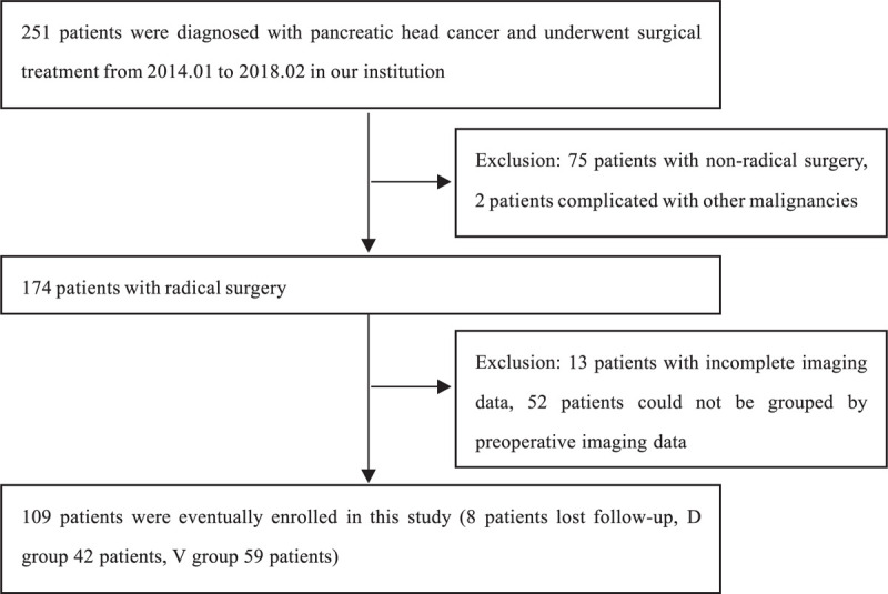
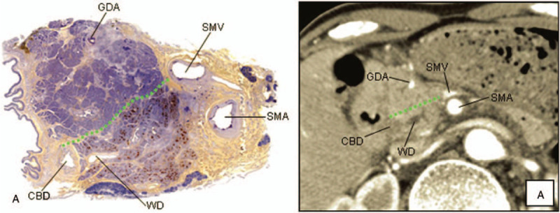
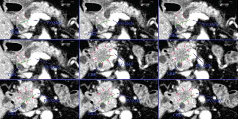
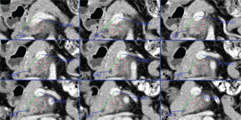
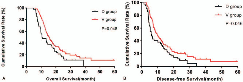
The embryonic development of the pancreas originates from dorsal and ventral anlagen, and the pancreatic cancer arising from dorsal or ventral pancreas may have different clinical pathology features. This study aims to explore whether there are differences in clinicopathological features and prognosis of pancreatic head carcinoma arising from dorsal or ventral pancreas.Between January 2014 and February 2018, 101 patients with resectable pancreatic head cancer who underwent pancreaticoduodenectomy in our institution were retrospectively reviewed. The patients were assigned into 2 groups according to tumor location on preoperative imaging materials (computed tomography/magnetic resonance imaging [CT/MRI]), and the clinicopathological features and prognosis were retrospectively analyzed in view of the embryonic development of the pancreas.Among these patients with pancreatic head cancer, 42 patients had tumors arising from dorsal pancreas (D group) and 59 patients had tumors arising from ventral pancreas (V group). The frequency of lymph node (LN) metastasis around the common hepatic artery (CHA) and hepatoduodenal ligament lymph nodes in the D group was higher than that in the V group (45.2% vs 10.2%, P = .001). And the rate of LN metastasis in the superior mesenteric artery (SMA) region in the V group is higher than that in the D group (32.2% vs 4.8%, P = .002). The D group was more likely to invade the common bile duct (78.6% vs 59.3%, P = .042) and duodenum (71.4% vs 44.1%, P = .006) than the V group. In addition, the survival outcome of V group was better than D group (median overall survival [OS], 15.37 months vs 10.53 months, P = .048, median DFS 9.73 months vs 5.93 months, P = .046).The clinicopathological features of pancreatic head carcinoma arising from dorsal or ventral pancreas are different, and the pancreatic head carcinoma arising from ventral pancreas has a better survival outcome.
Copyright © 2021 the Author(s). Published by Wolters Kluwer Health, Inc.
Conflict of interest statement
The authors have no conflicts of interest to disclose.
Figures
References
-
- Siegel RL, Miller KD, Jemal A. Cancer statistics, 2018. CA Cancer J Clin 2018;68:07–30. - PubMed
-
- Tempero MA, Malafa MP, Al-Hawary M, et al. . Pancreatic adenocarcinoma, Version 2.2017. J Natl Compr Canc Netw 2017;15:1028–61. - PubMed
-
- Tempero MA, Malafa MP, Behrman SW, et al. . Pancreatic adenocarcinoma, Version 2.2014 featured updates to the NCCN Guidelines. J Natl Compr Canc Netw 2014;12:1083–93. - PubMed
-
- Hartwig W, Werner J, Jaeger D, et al. . Improvement of surgical results for pancreatic cancer. Lancet Oncol 2013;14:E476–85. - PubMed
Publication types
MeSH terms
LinkOut - more resources
Full Text Sources
Medical
