. 2021 Jun 4;18(1):116.
doi: 10.1186/s12985-021-01579-z.

Lithium inhibits NF-κB nuclear translocation and modulate inflammation profiles in Rift valley fever virus-infected Raw 264.7 macrophages

Affiliations

Lithium inhibits NF-κB nuclear translocation and modulate inflammation profiles in Rift valley fever virus-infected Raw 264.7 macrophages

Raymond Tshepiso Makola et al. Virol J. .

Abstract

Introduction: Rift Valley fever virus (RVFV) is a zoonotic life-threatening viral infection endemic across sub-Saharan African countries and the Arabian Peninsula; however, there is a growing panic of its spread to non-endemic regions. This viral infection triggers a wide spectrum of symptoms that span from fibril illnesses to more severe symptoms such as haemorrhagic fever and encephalitis. These severe symptoms have been associated with dysregulated immune response propagated by the virulence factor, non-structural protein (NSs). Thus, this study investigates the effects of lithium on NF-κB translocation and RFVF-induced inflammation in Raw 264.7 macrophages.

Methods: The supernatant from RVFV-infected Raw 264.7 cells, treated with lithium, was examined using an ELISA assay kit to measure levels of cytokines and chemokines. The H2DCF-DA and DAF-2 DA florigenic assays were used to determine the levels of ROS and RNS by measuring the cellular fluorescence intensity post RVFV-infection and lithium treatment. Western blot and immunocytochemistry assays were used to measure expression levels of the inflammatory proteins and cellular location of the NF-κB, respectively.

Results: Lithium was shown to stimulate interferon-gamma (IFN-γ) production as early as 3 h pi. Production of the secondary pro-inflammatory cytokine and chemokine, interleukin-6 (IL-6) and regulated on activation, normal T cell expressed and secreted (RANTES), were elevated as early as 12 h pi. Treatment with lithium stimulated increase of production of tumor necrosis factor-alpha (TNF-α) and Interleukin-10 (IL-10) in RVFV-infected and uninfected macrophages as early as 3 h pi. The RVFV-infected cells treated with lithium displayed lower ROS and RNS production as opposed to lithium-free RVFV-infected control cells. Western blot analyses demonstrated that lithium inhibited iNOS expression while stimulating expression of heme oxygenase (HO) and IκB in RVFV-infected Raw 264.7 macrophages. Results from immunocytochemistry and Western blot assays revealed that lithium inhibits NF-κB nuclear translocation in RVFV-infected cells compared to lithium-free RVFV-infected cells and 5 mg/mL LPS controls.

Conclusion: This study demonstrates that lithium inhibits NF-kB nuclear translocation and modulate inflammation profiles in RVFV-infected Raw 264.7 macrophage cells.

Keywords: Inflammation; Macrophages; RVFV and NF-kB.


Conflict of interest statement

There are no financial and non-financial competing interests.

Figures

Fig. 1
Fig. 1
Determination of the effects lithium on TNF-α production after inoculation with the RVFV. In order to measure effects of lithium in TNF-α, IL-6, IL-10, IFN-γ and RANTES production, Raw 264.7 cells were seeded at the 2.4 × 10⁶ cells per T 25 flask for 3 h inoculated with 1 × 104.8 viral titre/mL for an hour. The excess virus was washed and the cells were treated with various concentration of lithium and then the supernatant was collected in 3, 6, 12 and 24 h time intervals. The Sandwich Elisa (PeproTech, USA) was executed to measure the amount of these cytokines from various time points. The data points represent the mean + standard deviation (error bar). The plot was developed with Graph-Pad Prism-6 software and GraphPad InStat-3 was used to establish the statistical analysis
Fig. 2
Fig. 2
Determination of the effects of lithium on oxidative burst after Raw 264.7 cell are challenged with RVFV and expression of levels of the antioxidant enzyme. a Cells were seeded at 4 × 10⁵ cells/well in a 6 well plate for 3 h and then inoculated with 1 × 104.8 viral titre/mL for an hour, the excess virus was substituted with fresh media and various lithium concentrations as well as 5 mg/mL LPS for 24 h. After 24 h of inoculation, cells were stained with H2DCF-DA at RT for 30 min in the dark and then cell fixed with 3.7% paraformaldehyde for an hour. The pictures where captured with EVOS FL Colour imaging system (Life Technologies, USA) Ex: 495 nm; Em: 515 nm. b Cells were seeded at 1 × 10⁶ cells/well in a 96 well plate for 3 h and then inoculated with RVFV at 103.8 viral titter/100uL for an hour. The excess virus was then substituted with fresh media and lithium concentrations as well as 5 mg/mL LPS, this was incubated for 12 and 24 h. After the incubation hours the cells were stained with H2DCF-DA for 30 min in the dark, and then the fluorescence intensity was measured with Fluoroskan Ascent FL (Thermo Fisher Scientific, USA) at ex (485 nm)-em (538 nm). The graphs were developed with Graph Pad Prism-6 software and GraphPad InStat-3 was used to establish the statistical analysis. c In order to determine the expression of HO-1 protein, Raw 264.7 cell where seeded at 1 × 10⁶ cell/mL for 3 h and then inoculated with 104.8 viral titre/mL for 1 h, and then the excess virus was substituted with a fresh media and lithium concentrations for 12 h. This was then followed by isolation of proteins and then western blotting assay. The pictures were captured with ChemiDoc XRS+ (Bio-RAD, USA)
Fig. 3
Fig. 3
Determination of the effects of lithium on the production of inflammatory reactive nitrogen species 24 h post RVFV inoculation and expression levels of the NOS enzyme. a Cells were seeded at 4 × 10⁵ cells/well in a 6 well plate for 3 h and then inoculated with 1 × 104.8 viral titre/mL for an hour, the excess virus was substituted with fresh media and various lithium concentrations as well as 5 mg/mL LPS for 24 h. After 24 h of inoculation, cells were staining with DAF-2 DA at RT for 30 min in the dark then cell were fixed with 3.7% paraformaldehyde for an hour. The pictures where captured with EVOS FL Colour imaging system (Life technologies, USA). b Cells were seeded at 1 × 10⁶ cells/well for 3 h and then inoculated with RVFV at 103.8 viral titter/100 μL for an hour. The excess virus was then substituted with fresh media and lithium concentrations as well as 5 mg/mL LPS for 12 and 24 h. After the incubation hours the cells were stained with DAF-2 DA for 30 min in the dark, and then the fluorescence was measured with Fluoroskan Ascent FL (Thermo Fisher Scientific, USA) at ex (485 nm)-em (538 nm). The plot were developed with Graph Pad Prism-6 software and GraphPad InStat-3 was used to establish the statistical analysis. c In order to determine the expression of NOS-2 protein, Raw 264.7 cell were seeded at 1 × 10⁶ cell/mL for 3 h and then inoculated with 104.8 viral titre/mL for 1 h, and then the excess virus was substituted with a fresh media and lithium concentrations for 12 h. This was then followed by isolation of proteins and then western blotting assay. The pictures were captured with ChemiDoc XRS+ (Bio-RAD, USA)
Fig. 4
Fig. 4
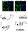
Effects of lithium on translocation of NF-kB between the cytoplasm and nucleus, and expression levels of IkB. a The cells were seeded at 4 × 10⁵ cells/well in a 6 well plate for 3 h and then inoculated with 1 × 104.8 viral titre/mL for an hour, the excess virus was substituted with fresh media and various lithium concentrations as well as 5 mg/mL LPS for 24 h. After 24 h of inoculation, cells were fixed with 4% paraformaldehyde for an hour, and then permeabilised with (0.1% Triton X-100, 1%BSA) for 30 min. Permeabilised cells were incubated for 60 min with rabbit anti-p65 antibody (1:500). The primary antibody was followed by FITC-labelled goat anti-rabbit secondary Ab incubation for 60 min. Thereafter, nuclear staining with 25µg/mL DAPI was executed for 5min in the dark. Cells were mounted on slides using 50% glycerol and pictures were captured with fluorescent inverted Nikon Ti-E microscope at 20× magnification. b and c In order to determine the translocation quantity of the NF-κB, Raw 264.7 cell were seeded at 1 × 10⁶ cell/mL for 3 h in the T25 cell culture flasks and then inoculated with 1 × 104.8 viral titre/mL for an hour, the excess virus was substituted with fresh media and various lithium concentrations as well as 5 mg/mL LPS for 24 h. This was then followed by isolation of cytoplasmic proteins as well the nucleus proteins and then western blotting assay followed. The pictures were captured with ChemiDoc XRS+ (Bio-RAD, USA). The ChemiDoc XRS+ image lab 5.2.1 software was used to measure band volume (Bio-RAD, USA). c The plot was developed with Graph pad prism-6 software and instat-3 was used to establish the statistical analysis. d In order to determine the expression of Ikb-α protein, Raw 264.7 cell were seeded at 1 × 10⁶ cell/mL for 3 h and then inoculated with 104.8 viral titre/mL for 1 h, and then the excess virus was substituted with a fresh media and lithium concentrations for 12 h. This was then followed by isolation of proteins and then western blotting assay. The ChemiDoc XRS+ image lab 5.2.1 software was used to capture pictures and measure band volume (Bio-RAD, USA)
Fig. 5
Fig. 5
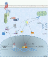
Determination of the canonical NF-kB and IRF3/7 signalling pathways and the effects of lithium post RVFV infection. TLR 2 and 4 are stimulated by the viral glycoproteins that in turn recruit adapter molecule Myd88 via tirap. The adapter molecules recruit Irak4 which phosphorylate recruit irak-1 which then associate with Traf-6. Traf-6 recruit Tak1 and Tab2. Tak1 phosphorylate IΚΚ-β, which then phosphorylate IkB which is then tagged for ubiquitination and then degradation by cytoplasmic proteases. This then allows translocation of NF-kB to the nucleus and inflammatory genes expression. The RIG-1 is known to be stimulated by dsRNA from replicating viral genome, which is said to be hidden from the TLR-3. This cytoplasmic receptor is shown to be essential for viral IFN expression. The RIG-1 is shown to associate with IPS-1 with its N-terminal card domain. The IPS-1 and RIG-1 association activate TBK1 and IΚΚ-ε which phosphorylate IRF-3 and 7. The NSs is suggested to interfere with the IFN signalling at the transcription factor level since there is an expression of other inflammatory mediators except for IFNs. Since, the NSs inhibit the interferon production via IRF inhibition other transcription factors such NF-kB continue to produce inflammatory mediators, hence, elevated production of other inflammatory mediators except the IFN. This diagram suggests that NF-kB inhibition as a result of upregulated IkB could be the, mechanism in which lithium restore dysregulated inflammation after RVFV infection leading to haemorrhagic fever pathogeneses observed during this viral infection

Similar articles

Cited by

References

    1. Akira S, Uematsu S, Takeuchi O. Pathogen recognition and innate immunity. J Cell. 2006;124:783–801. doi: 10.1016/j.cell.2006.02.015. - DOI - PubMed
    1. Caroline AL, Kujawa MR, Oury TD, Reed DS, Hartman AL. Inflammatory biomarker associated with Rift Valley fever encephalitis in the Lewis Rat model. Front Microbiol. 2016;6:1509. doi: 10.3389/fmicb.2015.01509. - DOI - PMC - PubMed
    1. Christen Y. Oxidative stress and Alzheimer’s disease. Am J Clin Nutr. 2000;71:621–629. doi: 10.1093/ajcn/71.2.621s. - DOI - PubMed
    1. Garcia MA, Gallego P, Campagna M, Gonzalez-Santamarıa J, Martınez G. Activation of NF-kB pathway by virus infection requires Rb expression. PLoS ONE. 2009;4(7):e6422. doi: 10.1371/journal.pone.0006422. - DOI - PMC - PubMed
    1. Ghaemi-Bafghi M, Haghparast A. Viral evasion and subversion mechanisms of the host immune system. ZJRMS. 2013;15(10):6–1.

Publication types