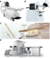
Image-Guided High-Intensity Focused Ultrasound, A Novel Application for Interventional Nuclear Medicine?
- PMID: 34088775
- PMCID: PMC8882895
- DOI: 10.2967/jnumed.120.256230
Image-Guided High-Intensity Focused Ultrasound, A Novel Application for Interventional Nuclear Medicine?
Abstract
Image-guided high-intensity focused ultrasound (HIFU) has been increasingly used in medicine over the past few decades, and several systems for such have become commercially available. HIFU has passed regulatory approval around the world for the ablation of various solid tumors, the treatment of neurologic diseases, and the palliative management of bone metastases. The mechanical and thermal effects of focused ultrasound provide a possibility for histotripsy, supportive radiation therapy, and targeted drug delivery. The integration of imaging modalities into HIFU systems allows for precise temperature monitoring and accurate treatment planning, increasing the safety and efficiency of treatment. Preclinical and clinical results have demonstrated the potential of image-guided HIFU to reduce adverse effects and increase the quality of life postoperatively. Interventional nuclear image-guided HIFU is an attractive noninvasive option for the future.
Keywords: PET/MR image guidance; hyperthermia; image-guided HIFU; targeted drug delivery; tumor ablation.
© 2021 by the Society of Nuclear Medicine and Molecular Imaging.
Figures


Similar articles
-
Magnetic Resonance-Guided High-Intensity Focused Ultrasound (MR-HIFU): Technical Background and Overview of Current Clinical Applications (Part 1).Rofo. 2019 Jun;191(6):522-530. doi: 10.1055/a-0817-5645. Epub 2019 Jan 10. Rofo. 2019. PMID: 30630200 Review. English.
-
Evolution of the ablation region after magnetic resonance-guided high-intensity focused ultrasound ablation in a Vx2 tumor model.Invest Radiol. 2013 Jun;48(6):381-6. doi: 10.1097/RLI.0b013e3182820257. Invest Radiol. 2013. PMID: 23399810
-
Interleaved Mapping of Temperature and Longitudinal Relaxation Rate to Monitor Drug Delivery During Magnetic Resonance-Guided High-Intensity Focused Ultrasound-Induced Hyperthermia.Invest Radiol. 2017 Oct;52(10):620-630. doi: 10.1097/RLI.0000000000000392. Invest Radiol. 2017. PMID: 28598900
-
Magnetic resonance guided high-intensity focused ultrasound for image-guided temperature-induced drug delivery.Adv Drug Deliv Rev. 2014 Jun;72:65-81. doi: 10.1016/j.addr.2014.01.006. Epub 2014 Jan 22. Adv Drug Deliv Rev. 2014. PMID: 24463345 Review.
-
Advances in MR image-guided high-intensity focused ultrasound therapy.Int J Hyperthermia. 2015 May;31(3):225-32. doi: 10.3109/02656736.2014.976773. Epub 2014 Nov 6. Int J Hyperthermia. 2015. PMID: 25373687 Review.
Cited by
-
Focused Ultrasound Treatment of a Spheroid In Vitro Tumour Model.Cells. 2022 Apr 30;11(9):1518. doi: 10.3390/cells11091518. Cells. 2022. PMID: 35563823 Free PMC article.
-
Ultrasound-induced cavitation renders prostate cancer cells susceptible to hyperthermia: Analysis of potential cellular and molecular mechanisms.Front Genet. 2023 Apr 19;14:1122758. doi: 10.3389/fgene.2023.1122758. eCollection 2023. Front Genet. 2023. PMID: 37152995 Free PMC article.
-
Real-Time and Delayed Imaging of Tissue and Effects of Prostate Tissue Ablation.Curr Urol Rep. 2023 Oct;24(10):477-489. doi: 10.1007/s11934-023-01175-4. Epub 2023 Jul 8. Curr Urol Rep. 2023. PMID: 37421582 Review.
-
Rational Nanomedicine Design Enhances Clinically Physical Treatment-Inspired or Combined Immunotherapy.Adv Sci (Weinh). 2022 Oct;9(29):e2203921. doi: 10.1002/advs.202203921. Epub 2022 Aug 24. Adv Sci (Weinh). 2022. PMID: 36002305 Free PMC article. Review.
References
-
- Kennedy JE. High-intensity focused ultrasound in the treatment of solid tumours. Nat Rev Cancer. 2005;5:321–327. - PubMed
-
- Siedek F, Yeo SY, Heijman E, et al. . Magnetic resonance-guided high-intensity focused ultrasound (MR-HIFU): technical background and overview of current clinical applications (part 1). Rofo. 2019;191:522–530. - PubMed
Publication types
MeSH terms
LinkOut - more resources
Full Text Sources
Other Literature Sources
Miscellaneous
