. 2021 Jun 8;118(23):e2022704118.
doi: 10.1073/pnas.2022704118.

Molecular basis of the dual role of the Mlh1-Mlh3 endonuclease in MMR and in meiotic crossover formation

Affiliations

Molecular basis of the dual role of the Mlh1-Mlh3 endonuclease in MMR and in meiotic crossover formation

Jingqi Dai et al. Proc Natl Acad Sci U S A. .

Abstract

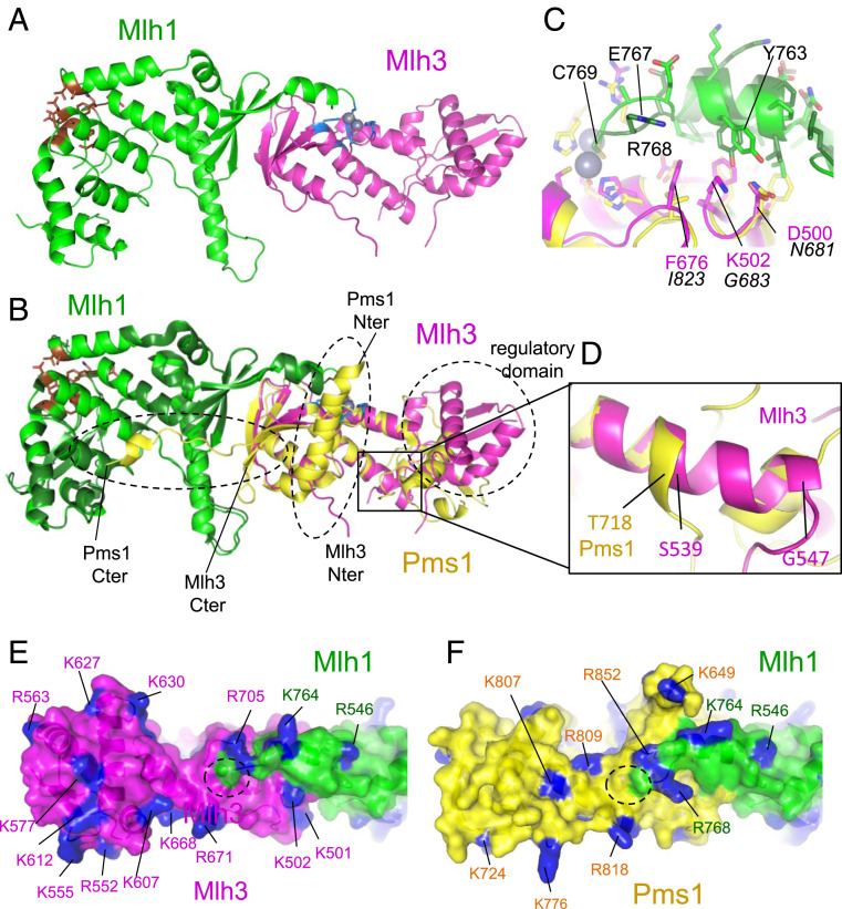
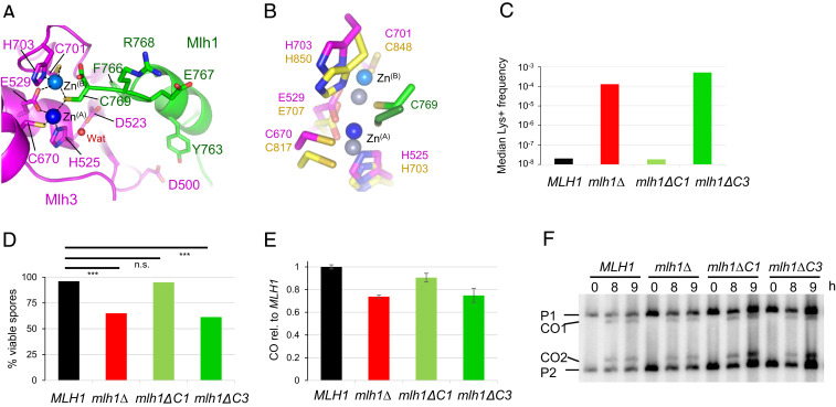
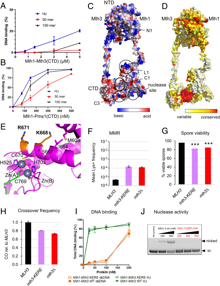
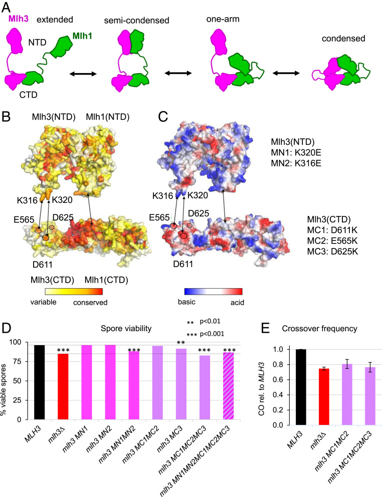
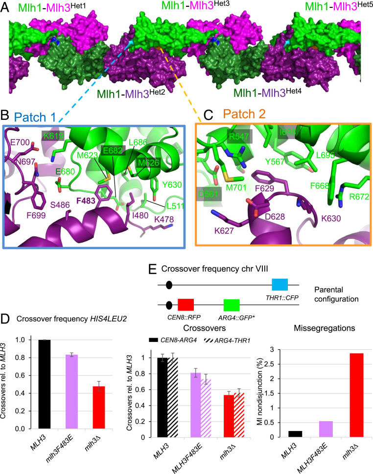
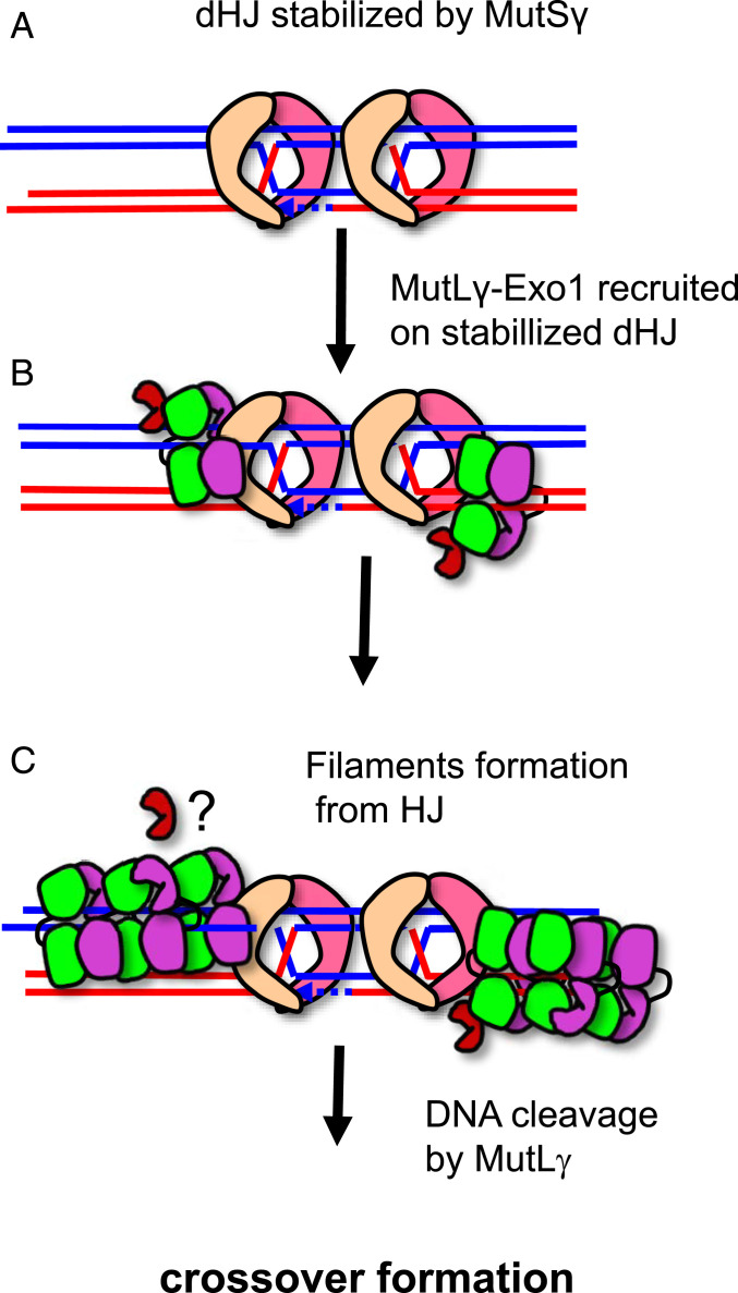
In budding yeast, the MutL homolog heterodimer Mlh1-Mlh3 (MutLγ) plays a central role in the formation of meiotic crossovers. It is also involved in the repair of a subset of mismatches besides the main mismatch repair (MMR) endonuclease Mlh1-Pms1 (MutLα). The heterodimer interface and endonuclease sites of MutLγ and MutLα are located in their C-terminal domain (CTD). The molecular basis of MutLγ's dual roles in MMR and meiosis is not known. To better understand the specificity of MutLγ, we characterized the crystal structure of Saccharomyces cerevisiae MutLγ(CTD). Although MutLγ(CTD) presents overall similarities with MutLα(CTD), it harbors some rearrangement of the surface surrounding the active site, which indicates altered substrate preference. The last amino acids of Mlh1 participate in the Mlh3 endonuclease site as previously reported for Pms1. We characterized mlh1 alleles and showed a critical role of this Mlh1 extreme C terminus both in MMR and in meiotic recombination. We showed that the MutLγ(CTD) preferentially binds Holliday junctions, contrary to MutLα(CTD). We characterized Mlh3 positions on the N-terminal domain (NTD) and CTD that could contribute to the positioning of the NTD close to the CTD in the context of the full-length MutLγ. Finally, crystal packing revealed an assembly of MutLγ(CTD) molecules in filament structures. Mutation at the corresponding interfaces reduced crossover formation, suggesting that these superstructures may contribute to the oligomer formation proposed for MutLγ. This study defines clear divergent features between the MutL homologs and identifies, at the molecular level, their specialization toward MMR or meiotic recombination functions.

Keywords: DNA recombination; DNA repair; biochemistry; genetics; structural biology.


Conflict of interest statement

The authors declare no competing interest.

Figures

Fig. 1.
Fig. 1.
Crystal structure of MutLγ(CTD). (A) Overall view of the MutLγ(CTD) heterodimer (Mlh1 and Mlh3 are colored respectively in green and magenta). The endonuclease site is colored in blue with the two zinc atoms in sphere representation and the Exo1 binding site in brown. (B) Superimposition of the MutLα(CTD) on the MutLγ(CTD). Mlh1 and Pms1 in MutLα are colored respectively in dark green and yellow. The three main regions of Pms1(CTD) that differ from Mlh3(CTD) are highlighted with dashed lines: 1) the regulatory domains, 2) the first residues of Pms1 and Mlh3 CTDs (the position of the first residues of the two CTDs are indicated [“Nter”]), and 3) the last residues of both CTDs (“Cter”). (C) The last residue Cys769 of Mlh1 adopts the same position in the endonuclease site in both complexes. The preceding residues of Mlh1 adopt a slightly different conformation when they are in contact with Mlh3 residues (numbers in magenta) or Pms1 ones (numbers in italic). (D) The helix αA of Mlh3 located in the hinge between the regulatory and dimerization domains is two turns longer in Mlh3 than in Pms1 pushing away the regulatory domain of Mlh3 compared to Pms1. The region zoomed corresponds to the black rectangle shown in B. The distribution of the basic residues (colored in blue) around the Mlh3 (E) and Pms1 (F) active sites is different (active site located with dashed lines). The position of the regulatory domain in Mlh3 also differs from the one observed in Pms1 resulting in different shapes of the surface surrounding the endonuclease sites of Mlh1-Mlh3 and Mlh1-Pms1.
Fig. 2.
Fig. 2.
Role of the last residues of Mlh1 in the active site of Mlh3 and Pms1. (A) Endonuclease site of MutLγ with two zinc atoms. Mlh1 and Mlh3 are colored respectively in green and magenta. The Zn atom called ZnA is colored in dark blue and the second one, called ZnB, is colored in light blue. Water molecules are colored in red. (B) Superimposition of Pms1 and Mlh3 active sites. ZnA and ZnB in Pms1 are colored in greys. Names of Pms1 residues are in yellow. C769 of Mlh1 in Mlh1-Pms1 complex is in green. (C) Mutation rates measured with a Lys+ reported assay with mlh1 alleles deleted of the last residue (mlh1∆C1) or the last three residues of Mlh1 (mlh1∆C3). The median values from a fluctuation analysis (see Materials and Methods) are plotted. MLH1 and mlh1∆ data were measured in (48). (D) Spore viability of diploid strains bearing the indicated MLH1 genotype at its endogenous locus. ***P < 0.001, Fisher’s exact test. Refer also to SI Appendix, Table S2. (EF) Crossing over frequency at the HIS4LEU2 hotspot monitored by Southern blot. Graph shows quantification at 8 and 9 h from two independent biological replicates ± range and are expressed relative to levels in MLH1 (same strains as in E).
Fig. 3.
Fig. 3.
DNA-binding properties of MutLγ and identification of a mlh3-KERE separation of function mutant. (A) EMSA of MutLγ(CTD) with a HJ with four arms of 25 bp each, 50 bp–long dsDNA or 100 bp–long dsDNA. The quantification of the gels is from the following number of experiments: HJ (n = 3), 50mer (n = 3), and 100mer (n = 2). Values are the mean ± SEM when n = 3, the mean ± range when n = 2. (B) EMSA of Mlh1-Pms1(CTD) with the same DNA substrates as in A. Values are the mean ± SEM from four independent experiments for each condition. (C and D) Molecular modeling of full-length Mlh1-Mlh3. The surface are colored according (C) to the electrostatic potential and (D) to the conservation rate of the amino acids deduced from multiple sequence alignments of Mlh1 or Mlh3 eukaryotes sequences. The circles represent the five main DNA-binding sites proposed from the literature and the experiments presented in this study. The N1, L1, and C1 sites are respectively in the NTD, linker, and CTD of Mlh1. The C3 is in the CTD of Mlh3. (E) The C3 site contains two basic residues (K668 and R671) that are exposed at the surface of Mlh3(CTD). These residues are close to the C670 position that is involved in the endonuclease site. (F) Mutation rate as measured with the Lys+ reporter assay of the Mlh3-KERE allele compared to wild type and mlh3Δ. Values are the mean of nine independent colonies ± SEM. (G) Spore viability of diploid strains bearing the indicated MLH3 genotype at its endogenous locus. ***P < 0.001, Fisher’s exact test. Refer also to SI Appendix, Table S2. (H) Crossing over frequency at the HIS4LEU2 hotspot monitored by Southern blot. Graph shows quantification from two independent biological replicates ± range and are expressed relative to levels in MLH3. (I) EMSA of wild-type Mlh1-Mlh3 and Mlh1-Mlh3(KERE) mutant with dsDNA and HJ. The quantification of the EMSA is from two independent experiments. Values are the mean ± range. (J) Nuclease activity of wild-type Mlh1-Mlh3 and Mlh1-Mlh3(KERE) mutant on supercoiled pUC19 plasmid DNA.
Fig. 4.
Fig. 4.
Condensation of Mlh1 and Mlh3 through NTD and CTD interactions. (A) Schematic representation of the four conformational states proposed for MutL homologs from AFM studies. (B and C) Surface representation of the NTD and CTD of Mlh1 and Mlh3 colored according to conservation rate of the amino acids (B) and to electrostatic potential (C). Two conserved basic residues in Mlh3-NTD (K316 and K320) and three conserved acid residues in Mlh3-CTD (E565, D611 and D625) are proposed to contribute to the condensation of the full-length heterodimer through their interaction in the condensed state. The five corresponding mutations in Mlh3(NTD) (MN1 and MN2) and in Mlh3(CTD) (MC1, MC2, and MC3) performed in this study are indicated. The structure of Mlh1 and Mlh3 NTDs were modeled from the crystal structures of MutL NTDs. (D) Spore viability of diploid strains bearing the indicated mlh3 genotype at its endogenous locus. The dotted lines indicate MLH3 (WT) and mlh3∆ spore viability levels for comparison. ***P < 0.001, **P < 0.01, Fisher’s exact test. Refer also to SI Appendix, Table S2. (E) Crossing over frequency at the HIS4LEU2 hotspot monitored by Southern blot. Graph shows quantification from three independent biological replicates ± SD, except for mlh3MC1MC2 (two replicates, mean values ± range), and are expressed relative to levels in MLH3 (same strains as in D).
Fig. 5.
Fig. 5.
Formation of MutLγ filaments through Mlh3-Cter and Mlh1-Cter interactions. (A) Filament-like structures made by Mlh1-Mlh3 heterodimers in the crystals. The interactions between five heterodimers (Het1 to Het5) in the crystal are represented by alternating the color of the heterodimers with Mlh1-Mlh3 in (green-magenta) or in (dark green-purple). The positions of the three main patches that mediate these structures are shown with the central residues colored respectively in cyan (patch 1), in orange (patch 2), and in blue (patch 3, SI Appendix, Fig. S5A). (B) Patch 1 includes interactions between six Mlh3 residues and nine Mlh1 residues. Most Mlh1 residues belong to the MIP-binding site that is involved in the interaction of Mlh1 with the MIP-motifs observed in Exo1, Ntg2 and Sgs1 (27). The F483 position of Mlh3 is buried in the center of this Mlh1 pocket in a position similar to the aromatic of the MIP motifs of the Mlh1 partners (SI Appendix, Fig. S5 AC, for the superposition of the MIP motif of Exo1 with the positions of the Mlh3 residues). (C) Patch 2 includes interactions between four Mlh3 residues and eight Mlh1 residues. The Mlh1 side contains the R547 position that interacts in MutLα with a Pms1 C-terminal extension that is not present in Mlh3(CTD). (D) Crossing over frequency at the HIS4LEU2 hotspot monitored by Southern blot. Graph shows quantification at 8 and 9 h from two independent biological replicates ± range and are expressed relative to levels in MLH3. (E) Meiotic crossovers on chromosome VIII. Top: illustration of the fluorescent spore setup (60). Genetic distances are measured in the CEN8-ARG4 and ARG4-THR1 adjacent intervals and expressed relative to the MLH3 values. The MI nondisjunction frequency was measured from the same dataset as for genetic distances, as described previously (60). MLH3: 1898 tetrads; mlh3F483E: 1277 tetrads; and mlh3∆: 1463 tetrads. Error bars: SE.
Fig. 6.
Fig. 6.
Model for MutLγ mechanism in meiotic recombination. In meiotic recombination, MutLγ, in complex with Exo1, is recruited by MutSγ on dHJ intermediates (AB). Transient MutLγ polymerization is proposed to occur at this step leading to an incision of the DNA away from the dHJ (C). This polymerization is proposed to involve three patches including the MIP-binding site of Mlh1. Further studies should help to evaluate whether competition exists between Exo1 and Mlh3 for the Mlh1 MIP-binding site.

Similar articles

Cited by

References

    1. Hunter N., Meiotic recombination: The essence of heredity. Cold Spring Harb. Perspect. Biol. 7, a016618 (2015). - PMC - PubMed
    1. Zickler D., Kleckner N., Recombination, pairing, and synapsis of homologs during meiosis. Cold Spring Harb. Perspect. Biol. 7, a016626 (2015). - PMC - PubMed
    1. Keeney S., Spo11 and the formation of DNA double-strand breaks in meiosis. Genome Dyn. Stab. 2, 81–123 (2008). - PMC - PubMed
    1. Allers T., Lichten M., Differential timing and control of noncrossover and crossover recombination during meiosis. Cell 106, 47–57 (2001). - PubMed
    1. Hunter N., Kleckner N., The single-end invasion: An asymmetric intermediate at the double-strand break to double-Holliday junction transition of meiotic recombination. Cell 106, 59–70 (2001). - PubMed

Publication types

MeSH terms

LinkOut - more resources