Review
. 2021 Dec;10(2):289-311.
doi: 10.1007/s40119-021-00222-w. Epub 2021 Jun 4.

Multidisciplinary Approaches for Transthyretin Amyloidosis

Affiliations
Review

Multidisciplinary Approaches for Transthyretin Amyloidosis

Haruki Koike et al. Cardiol Ther. 2021 Dec.

Abstract

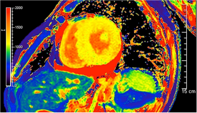
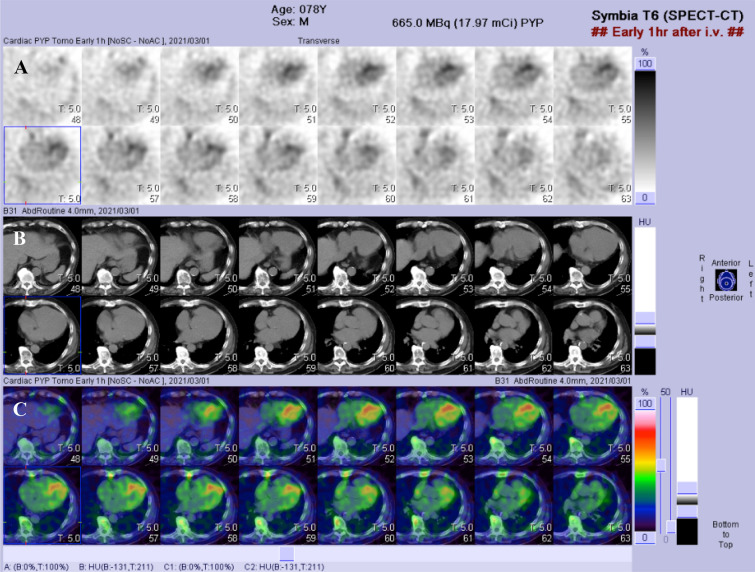
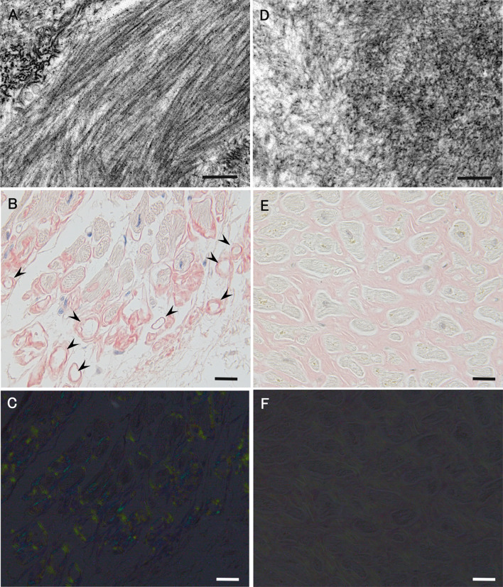
Amyloidosis caused by systemic deposition of transthyretin (TTR) is called ATTR amyloidosis and mainly includes hereditary ATTR (ATTRv) amyloidosis and wild-type ATTR (ATTRwt) amyloidosis. Until recently, ATTRv amyloidosis had been considered a disease in the field of neurology because neuropathic symptoms predominated in patients described in early reports, whereas advances in diagnostic techniques and increased recognition of this disease revealed the presence of patients with cardiomyopathy as a predominant feature. In contrast, ATTRwt amyloidosis has been considered a disease in the field of cardiology. However, recent studies have suggested that some of the patients with ATTRwt amyloidosis present tenosynovial tissue complications, particularly carpal tunnel syndrome, as an initial manifestation of amyloidosis, necessitating an awareness of this disease among neurologists and orthopedists. Although histopathological confirmation of amyloid deposits has traditionally been considered mandatory for the diagnosis of ATTR amyloidosis, the development of noninvasive imaging techniques in the field of cardiology, such as echocardiography, magnetic resonance imaging, and nuclear imaging, enabled nonbiopsy diagnosis of this disease. The mechanisms underlying characteristic cardiac imaging findings have been deciphered by histopathological studies. Novel disease-modifying therapies for ATTR amyloidosis, such as TTR stabilizers, short interfering RNA, and antisense oligonucleotides, were initially approved for ATTRv amyloidosis patients with polyneuropathy. However, the indications for the use of these disease-modifying therapies gradually widened to include ATTRv and ATTRwt amyloidosis patients with cardiomyopathy. Since the coronavirus disease 2019 (COVID-19) pandemic, which is caused by severe acute respiratory syndrome coronavirus 2 (SARS-CoV-2) infection, occurred, the minimization of hospital visits and telemedicine have become increasingly important. As older age and cardiovascular disease are major factors associated with increased disease severity and mortality of COVID-19, many ATTR amyloidosis patients are at increased risk of disease aggravation when they are infected with SARS-CoV-2. From this viewpoint, close interspecialty communication to determine the optimal interval of evaluation is needed for the management of patients with ATTR amyloidosis.

Keywords: Amyloid angiopathy; Cardiac amyloidosis; Inotersen; Patisiran; Positron emission tomography; Protein misfolding disease; Red flag; Scintigraphy; Tafamidis; Treatment.


Figures

Fig. 1
Fig. 1
Representative B-mode echocardiography findings of ATTR amyloidosis. a A thickened interventricular septum with hyperrefractile myocardial echoes (i.e., a granular sparkling appearance) is observed in the long-axis view (arrows). b Concentric hypertrophy of the ventricular wall is demonstrated in the short-axis view
Fig. 2
Fig. 2
Two-dimensional speckle-tracking strain imaging of echocardiography in a late-onset ATTRVal30Met amyloidosis patient from a nonendemic area of Japan. Reduced left ventricular longitudinal strain in the middle and basal segments with relatively preserved strain in the apex (i.e., apical sparing) is observed. A bull’s eye plot is shown in the lower right panel
Fig. 3
Fig. 3
Gadolinium-enhanced cardiac magnetic resonance imaging in a patient with ATTRwt amyloidosis. a Late gadolinium enhancement is observed along with the myocardial walls in an axial section. b The left ventricular wall shows diffuse subendocardial or transmural late gadolinium enhancement without a coronary distribution pattern
Fig. 4
Fig. 4
Microangiopathy in a late-onset ATTRVal30Met amyloidosis patient from a nonendemic area of Japan. A cross section of a sural nerve biopsy specimen. Uranyl acetate and lead citrate-stained specimens. The continuity of the endothelial cells of an endoneurial microvessel is lost (arrows), indicating the disruption of the blood–nerve barrier at this site. Scale bar 2 μm
Fig. 5
Fig. 5
Myocardial T1 mapping of magnetic resonance imaging in a late-onset ATTRVal30Met amyloidosis patient from a nonendemic area of Japan. Native myocardial T1 mapping indicates abnormally increased T1 relaxation time
Fig. 6.
Fig. 6.
99mTc-Pyrophosphate cardiac scintigraphy images in a late-onset ATTRVal30Met amyloidosis patient from a nonendemic area of Japan. a Myocardial uptake of the 99mTc-pyrophosphate tracer is observed on axial planes of single-photon emission computed tomography (SPECT). b Plain computed tomography (CT) images that correspond to a. c Reconstruction of SPECT images linked to plain CT images
Fig. 7
Fig. 7
Differential characteristics of amyloid deposits between early onset ATTRVal30Met amyloidosis patients from conventional endemic foci (ac) and late-onset ATTRVal30Met amyloidosis patients from nonendemic areas (df). Biopsy specimens of the sural nerve (a, d) and autopsy specimens of the heart (b, c, e, f). Uranyl acetate and lead citrate-stained specimens (a, d). Alkaline Congo red-stained specimens (b, c, e, f). On electron microscopy, amyloid fibrils tend to be long and thick in early onset patients from endemic foci (a). On light microscopy, amyloid deposits tend to be highly congophilic (b) and exhibit a strong apple-green birefringence under polarized light (c) in early onset patients from endemic foci. The atrophy and degeneration of myocardial cells result in the formation of amyloid rings (arrowhead). In late-onset patients from nonendemic areas, amyloid fibrils are generally short and thin on electron microscopy (d). On light microscopy, amyloid deposits are generally weakly congophilic (e) and exhibit a faint apple-green birefringence (f) in late-onset patients from nonendemic areas. Compared with (b), the atrophy of myocardial cells is not conspicuous despite massive amyloid deposition in (e). Scale bars 0.2 μm (a and d) and 20 μm (b, C, e, and f)

Similar articles

Cited by

References

    1. Adams D, Koike H, Slama M, Coelho T. Hereditary transthyretin amyloidosis: a model of medical progress for a fatal disease. Nat Rev Neurol. 2019;15(7):387–404. doi: 10.1038/s41582-019-0210-4. - DOI - PubMed
    1. Benson MD, Buxbaum JN, Eisenberg DS, Merlini G, Saraiva MJM, Sekijima Y, Sipe JD, Westermark P. Amyloid nomenclature 2020: update and recommendations by the International Society of Amyloidosis (ISA) nomenclature committee. Amyloid. 2020;27(4):217–222. doi: 10.1080/13506129.2020.1835263. - DOI - PubMed
    1. Koike H, Katsuno M. Transthyretin amyloidosis: update on the clinical spectrum, pathogenesis, and disease-modifying therapies. Neurol Ther. 2020;9(2):317–333. doi: 10.1007/s40120-020-00210-7. - DOI - PMC - PubMed
    1. Andrade C. A peculiar form of peripheral neuropathy; familiar atypical generalized amyloidosis with special involvement of the peripheral nerves. Brain. 1952;75(3):408–427. doi: 10.1093/brain/75.3.408. - DOI - PubMed
    1. Araki S, Mawatari S, Ohta M, Nakajima A, Kuroiwa Y. Polyneuritic amyloidosis in a Japanese family. Arch Neurol. 1968;18(6):593–602. doi: 10.1001/archneur.1968.00470360015001. - DOI - PubMed