Case Reports
. 2022 Aug;5(8):e1463.
doi: 10.1002/cnr2.1463. Epub 2021 Jun 5.

A case report of prostate cancer with leptomeningeal metastasis

Affiliations
Case Reports

A case report of prostate cancer with leptomeningeal metastasis

Mansoureh Dehghani et al. Cancer Rep (Hoboken). 2022 Aug.

Abstract

Background: Prostate cancer is the most prevalent cancer in men. However, leptomeningeal involvement by prostate carcinoma is a rare event.

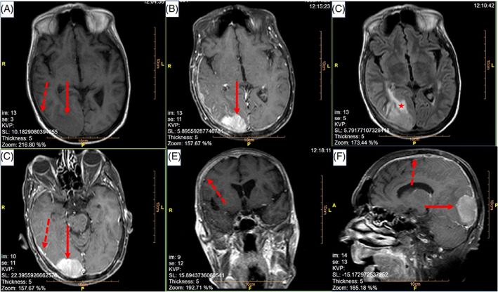
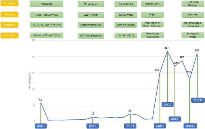
Case: Here, we report a 69-year-old patient with castration-resistant metastatic prostate cancer who presented with headache and ataxia. Brain MRI revealed a huge invasive interaxial mass at right occipital lobe with diffuse thickening and enhancement of meninges, the arachnoid, and the pia mater, and he was diagnosed with leptomeningeal carcinomatosis. The patient received whole brain radiotherapy.

Conclusion: Despite the fact that brain and leptomeningeal metastases are not very common in patients with prostate cancer, signs and symptoms of nervous system disorders should be assessed carefully, and consideration of such unusual metastases must be considered.

Keywords: leptomeningeal carcinomatosis; prostate cancer.


Conflict of interest statement

There was no conflict of interest to be reported.

Figures

FIGURE 1
FIGURE 1
MRI brain obtained from (A) axial plane, T1 without gadolinium contrast, (B) axial plane, T1 with contrast, (C) axial plane, T2, (D) axial plane, T1 with contrast, (E) coronal plane, T1 with contrast, and (F) sagittal plane, T1 with contrast. Solid lines show the 50 × 40 mm interaxial mass at right occipital lobe which was hypo/isosignal at T1‐weighted images and hypersignal at T2‐weighted images, with enhancement of the lesion at contrast‐enhanced T1‐weighted images. Dashed lines show diffuse leptomeningeal enhancement. The star shows the perilesional vasogenic edema
FIGURE 2
FIGURE 2
Clinical, biochemical, and treatment course of disease

References

    1. Wang N, Bertalan MS, Brastianos PK. Leptomeningeal metastasis from systemic cancer: review and update on management. Cancer. 2018;124(1):21‐35. 10.1002/cncr.30911. - DOI - PMC - PubMed
    1. Grossman SA, Krabak MJ. Leptomeningeal carcinomatosis. Cancer Treat Rev. 1999;25(2):103‐119. 10.1053/ctrv.1999.0119. - DOI - PubMed
    1. Orphanos G, Ardavanis A. Leptomeningeal metastases from prostate cancer: an emerging clinical conundrum. Clin Exp Metastasis. 2010;27(1):19‐23. 10.1007/s10585-009-9298-z. - DOI - PubMed
    1. Lin C, Turner S, Gurney H, et al. Increased detections of leptomeningeal presentations in men with hormone refractory prostate cancer: An effect of improved systemic therapy?. J Med Imaging Radiat Oncol. 2008;52(4):376‐381. 10.1111/j.1440-1673.2008.01973.x. - DOI - PubMed
    1. Neeman E, Salamon N, Rettig M. Leptomeningeal carcinomatosis of prostate cancer: a case report and review of the literature. Rev Urol. 2020;22(2):80‐84. - PMC - PubMed

Publication types