Review
. 2021 Jun 5;12(1):70.
doi: 10.1186/s13244-021-01006-5.

Computed tomography imaging of septic shock. Beyond the cause: the "CT hypoperfusion complex". A pictorial essay

Affiliations
Review

Computed tomography imaging of septic shock. Beyond the cause: the "CT hypoperfusion complex". A pictorial essay

Marco Di Serafino et al. Insights Imaging. .

Abstract

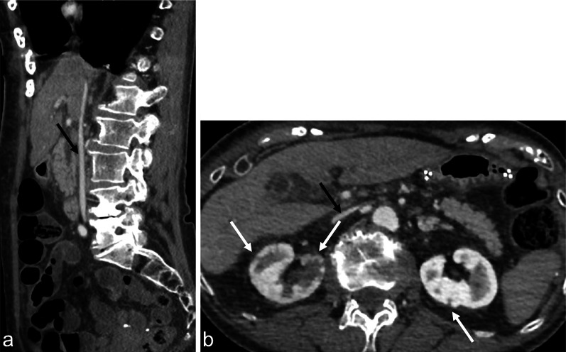
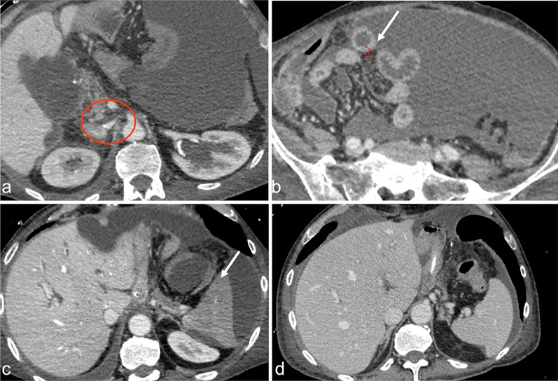
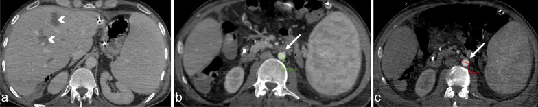
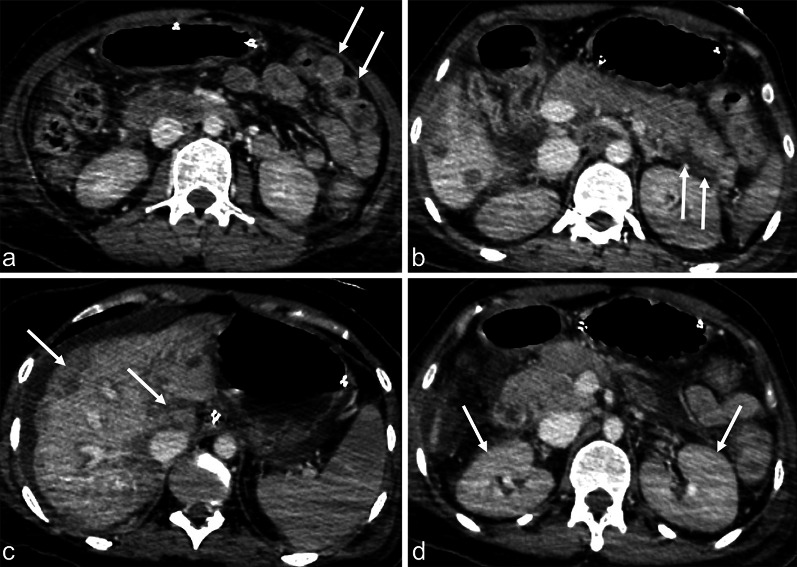
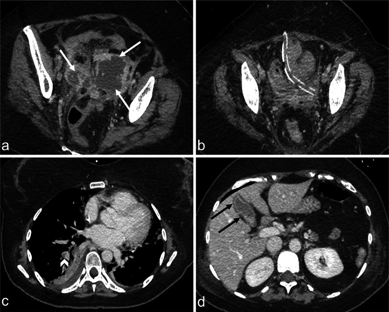
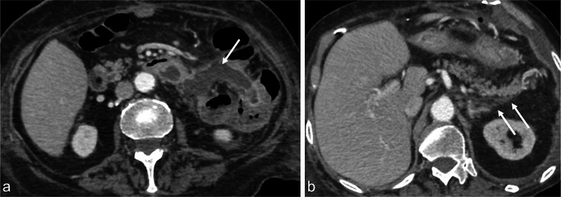
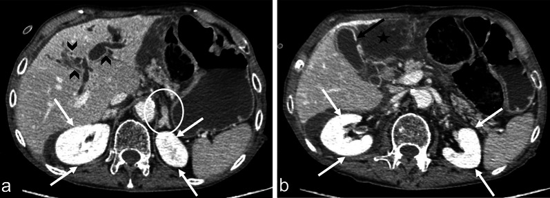
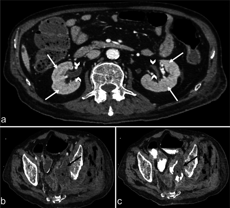
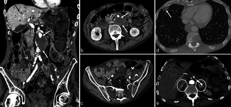
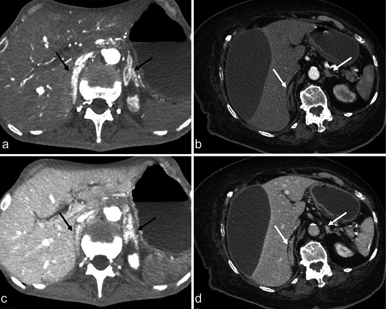
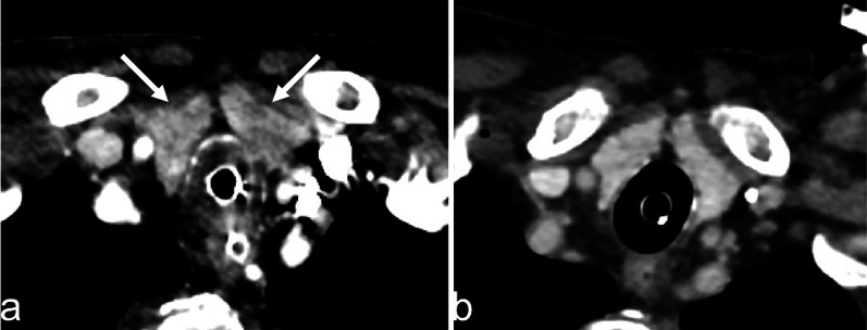
Septic shock is a medical emergency that represents one of the most important underlying causes for presentation to the Emergency Department. Sepsis is defined as organ dysfunction, life-threatening event caused by a deregulated inflammatory host response to infection, with a mortality risk ranging from 10 to 40%. Early sepsis identification is the cornerstone of management and diagnostic imaging can play a pivotal role in this clinical context. The choice of imaging modality depends on several factors, associated with the clinical condition and the presence or absence of localising signs and symptoms. The diagnostic accuracy of contrast-enhanced total-body CT has been well established during septic shock, allowing for a rapid, panoramic, and detailed study of multiple body areas, simultaneously. The aim of this article is to illustrate the controversial CT hypoperfusion complex in patients with septic shock, characterised by the following imaging features: decreased enhancement of the viscera; increased mucosal enhancement; luminal dilation of the small bowel; mural thickening and fluid-filled loops of the small bowel; the halo sign and flattening of the inferior vena cava; reduced aortic diameter; peripancreatic oedema; abnormal parenchymal perfusion; and other controversial findings that are variably associated with each other and reversible during the early stages. Increasing physicians' awareness of the significance of these findings could prompt alternative approaches to the early assessment and management of septic shock. In this perspective, CT imaging represents a useful tool for a complete, rapid and detailed diagnosis of clinically suspected septic shock, which can be used to improve patient outcomes.

Keywords: CT hypoperfusion complex; Contrast-enhanced CT; Diagnostic imaging; Sepsis; Septic shock.


Conflict of interest statement

The authors declare that they have no competing interests.

Figures

Fig. 1
Fig. 1
Contrast-enhanced CT in the portal venous phase, showing an 84-year-old male with sepsis (qSOFA 2) due to bilateral nephritis (white arrows). A collapsed IVC can be observed in both the sagittal (a, black arrow) and axial views (b, black arrow)
Fig. 2
Fig. 2
Contrast-enhanced CT in the portal venous phase, in the axial view, showing a 70-year-old male with sepsis due to gastric cancer surgery complications (qSOFA 3). In this patient, multiple CT signs of sepsis can be observed, including inferior vena cava halo sign (a, red circle), thickened bowel wall (b, white arrow), reduced enhancement, and spleen volume (c, white arrow). Figure d shows a previous CT examination of the same patient for comparison
Fig. 3
Fig. 3
Contrast-enhanced CT in the portal venous phase, in the axial plane, showing a 44-year-old female with sepsis (qSOFA 2) due to cholangitis with liver abscesses (a, arrowheads). The CT revealed the reduced calliper of the abdominal aorta (b, arrow), compared with the normal calliper of the abdominal aorta in a previous examination from the same patient (c)
Fig. 4
Fig. 4
Contrast-enhanced CT images in the portal venous phase, in the axial view, showing a 46-year-old female with lymphoma and sepsis due to infection (qSOFA 2). A thickened and hyperdense bowel wall can be observed (a, arrows), with peripancreatic oedema (b, arrows), reduced and inhomogeneous liver enhancement (c), and enlarged kidneys with abnormal enhancement (d, arrows)
Fig. 5
Fig. 5
Contrast-enhanced CT image in the portal venous phase showing a 44-year-old female with abscessed uterine neoplasm (a, arrows) and post-operative control with completely drained collection (b). Pulmonary thromboembolism (c, arrowhead) with development of a clinical septic state (qSOFA 2). Note the dense, gallbladder mural enhancement without thickened walls (d, black arrows)
Fig. 6
Fig. 6
Contrast-enhanced CT images in the portal venous phase, in the axial view, shows a 78-year-old female with sepsis (qSOFA 2) due to bowel anastomotic dehiscence (a, the white arrow indicates fluid collection caused by an anastomotic leak). Peripancreatic oedema can also be observed (b, white arrows)
Fig. 7
Fig. 7
Contrast-enhanced CT images in the portal venous phase, in the axial view, shows a 92-year-old male with sepsis of the biliary tract (qSOFA 3) characterised by segmental intrahepatic biliary duct dilatation (a, black arrowheads) and gallbladder leak (b, black arrow) with extrahepatic biloma (b, black star). Note the increased renal parenchymal enhancement bilaterally (a, b, white arrows) and the abnormal adrenal enhancement (a, white circle)
Fig. 8
Fig. 8
Contrast-enhanced CT images in the portal venous phase (a and b) and the three-minute-delayed phase (c), showing a patient with septic shock (qSOFA 2) due to entero-neovesical fistula (b and c, black arrows) with pyelitis (a, arrowheads) after radical cystectomy. The decreased enhancement of the renal medulla (a, white arrows) in the venous phase was observed
Fig. 9
Fig. 9
Contrast-enhanced CT images in the arterial phase showing a 75-year-old male with sepsis (qSOFA 3) due to an infected aneurysm after aorto-basilic stent placement (a and b, white arrows). Concomitant intestinal images reveal liver septic pneumatosis (a and b, black arrows), stercoraceous collection in the left iliac fossa (c, white arrowheads), and septic emboli with pulmonary infarction (d, white arrows). The adrenal glands display hyperenhancement (e, white circles)
Fig. 10
Fig. 10
Contrast-enhanced CT images in the arterial phase from two different patients with septic shock (a and b) and the respective venous phases (c and d). Different patterns of adrenal gland hyperenhancement can be observed, characterised in one patient by a central zone with less intense enhancement relative to the peripheral zone (a, black arrows) and in the other patient by a central zone with heterogeneous enhancement and a mosaic-like appearance (b, white arrows). In both cases, the venous phase shows a homogeneous enhancement (c, black arrows; d, white arrows)
Fig. 11
Fig. 11
Contrast-enhanced CT image in the portal venous phase showing an 89-year-old female with sepsis due to bowel wall perforation, secondary to colon cancer (qSOFA 3). An inhomogeneous enhancement of the thyroid gland was observed (a, arrow), in contrast with the previously homogeneous enhancement of the gland 4 days earlier (b)

Similar articles

Cited by

References

    1. Singer M, Deutschman CS, Seymour CW, et al. The third international consensus definitions for sepsis and septic shock (Sepsis-3) JAMA. 2016;315(8):801–810. doi: 10.1001/jama.2016.0287. - DOI - PMC - PubMed
    1. Dellinger RP, Levy MM, Rhodes A, et al. Surviving Sepsis Campaign: International guidelines for management of severe sepsis and septic shock 2012. Intensive Care Med. 2013;39(2):165–228. doi: 10.1007/s00134-012-2769-8. - DOI - PMC - PubMed
    1. Levy MM, Fink MP, et al. SCCM/ESICM/ACCP/ATS/SIS international sepsis definition conference. Intensive Care Med. 2001;29(4):530–538. doi: 10.1007/s00134-003-1662-x. - DOI - PubMed
    1. Shankar-Hari M, Phillips GS, Levy ML, Seymour CW, Liu VX, Deutschman CS, et al. Developing a new definition and assessing new clinical criteria for septic shock: for the third international consensus definitions for sepsis and septic shock (Sepsis-3) JAMA. 2016;315(8):775–787. doi: 10.1001/jama.2016.0289. - DOI - PMC - PubMed
    1. Levy MM, Evans LE, Rhodes A. The surviving sepsis campaign bundle: 2018 update. Intensive Care Med. 2018;44(6):925–928. doi: 10.1007/s00134-018-5085-0. - DOI - PubMed

LinkOut - more resources