

Impact of circulating miRNA-373 on breast cancer diagnosis through targeting VEGF and cyclin D1 genes
- PMID: 34089425
- PMCID: PMC8179880
- DOI: 10.1186/s43141-021-00174-7
Impact of circulating miRNA-373 on breast cancer diagnosis through targeting VEGF and cyclin D1 genes
Abstract
Background: Breast cancer (BC) is the common primary tumor among females. Hence, there is an urgent need to improve the early prediction and diagnosis of BC. For that reason, the object of the current study is to analyze the expression levels of miRNA-373 and its target genes including vascular endothelial growth factor (VEGF) and cyclin D1 in women with BC.
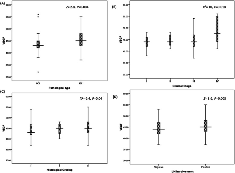
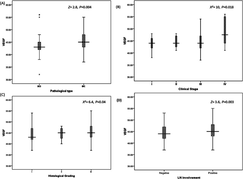
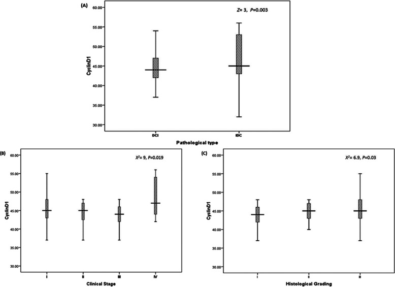
Results: Upregulation of miRNA-373 and its target genes was observed in BC patients followed by patients with benign breast lesions compared to downregulation in controls. There was a significant association between the expression level of miRNA-373 and all clinical features. The same associations were observed between its target genes and all clinico-pathological features except hormonal status. The correlation between miRNA-373 and both genes was significant.
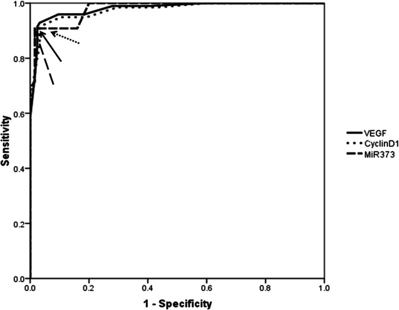
Conclusions: Our results prove that miRNA-373, as an oncomir, would be a vital biomarker for BC diagnosis and prognosis by targeting both VEGF and cyclin D1.
Keywords: Breast cancer; Clinico-pathological characteristics; Cyclin D1; Diagnosis; MicroRNA; VEGF.
Conflict of interest statement
The authors declare that they have no competing interests.
Figures
Similar articles
-
Role of COX-2, VEGF and cyclin D1 in mammary infiltrating duct carcinoma.Oncol Rep. 2003 Sep-Oct;10(5):1241-9. Oncol Rep. 2003. PMID: 12883688
-
Significance of Targeting VEGFR-2 and Cyclin D1 in Luminal-A Breast Cancer.Molecules. 2020 Oct 10;25(20):4606. doi: 10.3390/molecules25204606. Molecules. 2020. PMID: 33050377 Free PMC article.
-
[Expression of cyclin D1 and vascular endothelial growth factor(VEGF) in non-small cell lung carcinoma and their association with the prognosis].Ai Zheng. 2003 Jan;22(1):86-90. Ai Zheng. 2003. PMID: 12561444 Chinese.
-
Identification of Seven Cell Cycle-Related Genes with Unfavorable Prognosis and Construction of their TF-miRNA-mRNA regulatory network in Breast Cancer.J Cancer. 2021 Jan 1;12(3):740-753. doi: 10.7150/jca.48245. eCollection 2021. J Cancer. 2021. PMID: 33403032 Free PMC article.
-
The emerging role of miRNA clusters in breast cancer progression.Biochim Biophys Acta Rev Cancer. 2020 Dec;1874(2):188413. doi: 10.1016/j.bbcan.2020.188413. Epub 2020 Aug 20. Biochim Biophys Acta Rev Cancer. 2020. PMID: 32827583 Review.
Cited by
-
Regulation of Inflammasome by microRNAs in Triple-Negative Breast Cancer: New Opportunities for Therapy.Int J Mol Sci. 2023 Feb 7;24(4):3245. doi: 10.3390/ijms24043245. Int J Mol Sci. 2023. PMID: 36834660 Free PMC article. Review.
-
Targeting Triple-Negative Breast Cancer: Resistance Mechanisms and Therapeutic Advancements.Cancer Med. 2025 May;14(9):e70803. doi: 10.1002/cam4.70803. Cancer Med. 2025. PMID: 40318146 Free PMC article. Review.
-
MicroRNA Dysregulation in Early Breast Cancer Diagnosis: A Systematic Review and Meta-Analysis.Int J Mol Sci. 2023 May 5;24(9):8270. doi: 10.3390/ijms24098270. Int J Mol Sci. 2023. PMID: 37175974 Free PMC article.
-
Addressing the Clinical Feasibility of Adopting Circulating miRNA for Breast Cancer Detection, Monitoring and Management with Artificial Intelligence and Machine Learning Platforms.Int J Mol Sci. 2022 Dec 6;23(23):15382. doi: 10.3390/ijms232315382. Int J Mol Sci. 2022. PMID: 36499713 Free PMC article. Review.
-
Cell cycle associated miRNAs as target and therapeutics in lung cancer treatment.Heliyon. 2022 Oct 13;8(10):e11081. doi: 10.1016/j.heliyon.2022.e11081. eCollection 2022 Oct. Heliyon. 2022. PMID: 36303933 Free PMC article. Review.
References
LinkOut - more resources
Full Text Sources
Research Materials