. 2021 Jul;297(1):100851.
doi: 10.1016/j.jbc.2021.100851. Epub 2021 Jun 3.

Phosphorylation of human phospholipase A1 DDHD1 at newly identified phosphosites affects its subcellular localization

Affiliations

Phosphorylation of human phospholipase A1 DDHD1 at newly identified phosphosites affects its subcellular localization

Naoki Matsumoto et al. J Biol Chem. 2021 Jul.

Abstract

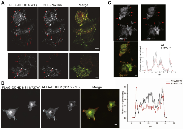
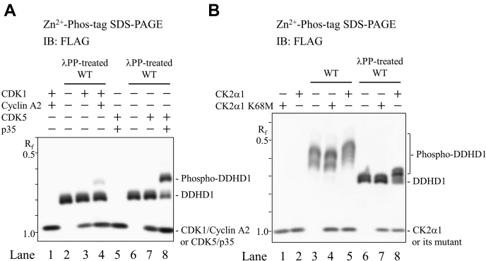
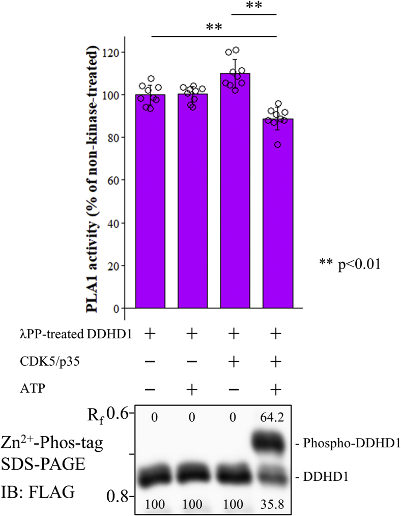
Phospholipase A1 (PLA1) hydrolyzes the fatty acids of glycerophospholipids, which are structural components of the cellular membrane. Genetic mutations in DDHD1, an intracellular PLA1, result in hereditary spastic paraplegia (HSP) in humans. However, the regulation of DDHD1 activity has not yet been elucidated in detail. In the present study, we examined the phosphorylation of DDHD1 and identified the responsible protein kinases. We performed MALDI-TOF MS/MS analysis and Phos-tag SDS-PAGE in alanine-substitution mutants in HEK293 cells and revealed multiple phosphorylation sites in human DDHD1, primarily Ser8, Ser11, Ser723, and Ser727. The treatment of cells with a protein phosphatase inhibitor induced the hyperphosphorylation of DDHD1, suggesting that multisite phosphorylation occurred not only at these major, but also at minor sites. Site-specific kinase-substrate prediction algorithms and in vitro kinase analyses indicated that cyclin-dependent kinase CDK1/cyclin A2 phosphorylated Ser8, Ser11, and Ser727 in DDHD1 with a preference for Ser11 and that CDK5/p35 also phosphorylated Ser11 and Ser727 with a preference for Ser11. In addition, casein kinase CK2α1 was found to phosphorylate Ser104, although this was not a major phosphorylation site in cultivated HEK293 cells. The evaluation of the effects of phosphorylation revealed that the phosphorylation mimic mutants S11/727E exhibit only 20% reduction in PLA1 activity. However, the phosphorylation mimics were mainly localized to focal adhesions, whereas the phosphorylation-resistant mutants S11/727A were not. This suggested that phosphorylation alters the subcellular localization of DDHD1 without greatly affecting its PLA1 activity.

Keywords: DDHD domain containing 1 (DDHD1); Phos-tag; cyclin-dependent kinase (CDK); hereditary spastic paraplegia (HSP); phospholipase A1 (PLA1); phosphorylation.


Conflict of interest statement

Conflict of interest The authors declare that they have no conflict of interest regarding the content of this article.

Figures

Figure 1
Figure 1
Human DDHD1 is a phosphorylated protein. Wild-type FLAG-DDHD1 (WT) and single (S8A, S11A, S723A, T726A, and S727A) or multiple (S8/11A, S11/727, S11/727A, S8/11/T726A, S8/11/727A, and S8/11/723/727A) Ser (Thr)-to-Ala substitution mutants were expressed in HEK293 cells. A, electrophoretic mobility shift in WT FLAG-DDHD1 by the inhibition of protein phosphatases. WT FLAG-DDHD1-expressing HEK293 cells were cultured in the absence (lane 1) or presence (lane 2) of 1 μM okadaic acid for 4 h in growth medium containing 10% FBS. Cell extracts were analyzed by Zn2+ Phos-tag SDS-PAGE following immunoblot analysis (IB). B, electrophoretic mobility shift in WT and Ala substitution mutants of human DDHD1 by phosphorylation. WT (lanes 1, 2, 8, and 9) and Ala substitution mutant (lanes 3–7 and 10–15)-expressing HEK293 cells were cultured in growth medium containing 10% FBS. Cell extracts were analyzed by Zn2+ Phos-tag SDS-PAGE (upper panels) or normal SDS-PAGE (lower panels) following immunoblot analysis (IB). In some cases, cell extracts from WT-expressing HEK293 cells were treated with λPP (lanes 1 and 8) to confirm electrophoretic mobility by phosphorylation. The Rf value of 1.0 is defined as the position of bromphenol blue dye. Results are from one experiment representative of three independent experiments.
Figure 2
Figure 2
Identification of phosphorylated sites in DDHD1 with or without okadaic acid. Extracts from FLAG-DDHD1-expressing and okadaic-acid-treated cells were immunoprecipitated with anti-DYKDDDDK (FLAG) antibody, fractionated by SDS-PAGE, and subjected to in-gel digestion with trypsin or AspN. The resultant peptides were analyzed by MALDI-TOF MS/MS. MS/MS spectra of a diphosphorylated peptide at Ser8 and Ser11 (A) and those at Ser723 and Thr726 (B) were depicted as representatives of the peptides shown in Table 1. The sequences of identified peptides are shown above the mass spectra; phosphorylation sites are indicated by PH on amino acids; bn denotes the ion generated by the cleavage of the peptide bond after the nth amino acid from the amino terminus; yn denotes the ions generated from the carboxyl terminus.
Figure 3
Figure 3
Effects of phosphorylation on the PLA1 activity of DDHD1.A, purified recombinant FLAG-DDHD1 (WT) was treated with or without λPP and PLA1 activity was measured using a fluorescent substrate (PED-A1). B and C, the PLA1 activities of WT, S8E, S11E, S723E, S727E, and S11/727E were measured after treatment without (B) or with λPP (C). Fifty nanograms of purified WT and mutants quantified by bicinchoninic acid assay were used as enzymes for these assays. All three experiments were normalized by the averaged value of WT (left) in combination. Results are expressed as the mean ± SD of three experiments performed in triplicate (n = 9). Each individual point is also represented as a small circle. Statistical analyses were performed by the Student’s t test (two asterisks, p < 0.01).
Figure 4
Figure 4
Localization of phosphorylated DDHD at focal adhesions.A, ALFA-DDHD1 (WT) and GFP-Paxillin were expressed in COS-7 cells on retronectin-coated coverglasses and the cells were immunostained with tdTomato-anti-ALFA tag nanobody. The fluorescent signals of ALFA tag (left, red) and GFP (right, green) were acquired. Images 1.5 to 2.5 μm away from the glass surface are shown. Typical focal contacts are indicated by arrows. Bar, 5 μm. B, FLAG-DDHD1 (S11/727A) and ALFA-DDHD1 (S11/727E) were coexpressed in COS-7 cells and stained with FLAG-tag antibody (red) and anti-ALFA nanobody labeled with cfSGFP2 (green). The phospho-mimic form (S11/727E) was enriched at regions of focal adhesion, which was not observed with the phosphorylation-resistant mutant (S11/727A). The intensity map at the indicated line (blue) is also shown. C, immunostained FLAG-DDHD1 (WT) and ALFA-DDHD1 S11/S727A. Focal contacts are marked with arrows and the signal at the line (blue) was quantified. Bar, 5 μm.
Figure 5
Figure 5
Effects of protein kinase inhibitors on the phosphorylation of DDHD1. FLAG-DDHD1-expressing PANC1 cells were cultured in the absence (lanes 1–3) or presence of 100 μM olomoucine (lane 4) or 10 (lane 5), or 100 μM CHIR99021 (lane 6) for 2 days with growth medium containing 10% FBS. Cell extracts from vehicle-treated cells were incubated with (lane 1) or without calf intestine alkaline phosphatase (CIAP) (lane 2). Cell extracts were analyzed by Zn2+ Phos-tag SDS-PAGE following immunoblot analysis (IB). The Rf value of 1.0 is defined as the position of bromphenol blue dye. Results are from one experiment representative of three independent experiments.
Figure 6
Figure 6
Phosphorylation of DDHD1 by CDK1, CDK5, and CK2α1.A, Phosphorylation by CDK1 or CDK5. The dephosphorylated form of purified recombinant FLAG-DDHD1 (λPP-treated WT, lanes 2–4 and 6–8) or TBS (none, lanes 1 and 5) was incubated in combination with purified FLAG-tagged CDK1 or CDK5, and their activators, cyclin A2 or p35, at 25 °C for 180 min. B, Phosphorylation by CK2α1, recombinant DDHD1 (WT, lanes 3–5), its dephosphorylated form (λPP-treated WT, lanes 6–8), or TBS (none, lanes 1 and 2) was incubated in combination with purified FLAG-tagged CK2α1 or its kinase-inactive form K68M at 25 °C for 180 min. The incubated mixtures containing equivalent amounts of DDHD1 were analyzed by Zn2+ Phos-tag SDS-PAGE following immunoblot analysis using anti-FLAG M2 antibody. The Rf value of 1.0 is defined as the position of bromphenol blue dye. Results are from one experiment representative of three independent experiments.
Figure 7
Figure 7
Identification of phosphorylation sites of DDHD1 targeted by CDK1, CDK5, and CK2α1.A and B, Phosphorylation by CDK1 or CDK5. The dephosphorylated form of purified recombinant FLAG-DDHD1 (WT) or its alanine mutants (S8/11A, S8/727A, S11/727A, S8/11/T726A, and S8/11/727A) was incubated with or without purified FLAG-tagged CDK1/cyclin A2 (A) or CDK5/p35 (B) at 25 °C for 150 min. C, Phosphorylation by CK2α1, the dephosphorylated form of purified recombinant FLAG-DDHD1 (WT) or its alanine mutants (S104A and S8/11/727A) was incubated with or without purified FLAG-tagged CK2α1 at 25 °C for 180 min. The incubated mixtures containing equivalent amounts of DDHD1 were analyzed by Zn2+ Phos-tag SDS-PAGE following immunoblot analysis using anti-FLAG M2 antibody. Blots were exposed to ECL reagent in order to detect up-shifted bands by CDK1/cyclin A2 or CDK5/p35. The Rf value of 1.0 is defined as the position of bromphenol blue dye. Results are from one experiment representative of three independent experiments.
Figure 8
Figure 8
Effects of CDK5/p35 on the PLA1 activity of DDHD1. The dephosphorylated form of purified recombinant FLAG-DDHD1 was incubated with or without purified FLAG-tagged CDK5/p35 or ATP at 25 °C for 360 min, and the PLA1 activity of reaction mixtures containing 50 ng of DDHD1 was compared. All three experiments normalized by the averaged value of WT without CDK5/p35 and ATP (left-most) are combined. Results are expressed as the mean ± SD of three experiments performed in triplicate (n = 9). Each individual point is also represented as a small circle. Statistical analyses were performed by the Student’s t test (two asterisks, p < 0.01). The lower panel shows phosphorylation states at the time of the PLA1 activity measurement detected by Zn2+ Phos-tag SDS-PAGE followed by immunoblot analysis (IB) using anti-FLAG M2 antibody, and the numeric characters below each band indicate the abundance ratio of DDHD1 and phosphorylated DDHD1. The Rf value of 1.0 is defined as the position of bromphenol blue dye.
Figure 9
Figure 9
Summary of the phosphorylation of human DDHD1, potential responsible protein kinases, and possible physiological function. The lipase consensus sequence and DDHD1 domain are shown in red and light orange, respectively. The glycine-rich and coiled-coil domains are shown in pale blue and green, respectively. Numbers, such as 1 and 872, are those of amino acid residues. The phosphorylation sites identified in the present study are depicted as P in yellow circles. The phosphorylation sites by CDK1/cyclin A2, CDK5/p35, and CK2α1 are indicated. Translocation of DDHD1 to focal adhesions by phosphorylation at Ser11 and Ser727 is depicted.

Similar articles

Cited by

References

    1. Waite M. vol 5. Plenum; New York, NY: 1987. pp. 1–332. (The Phospholipases: Handbook of Lipid Research).
    1. Pete M.J., Ross A.H., Exton J.H. Purification and properties of phospholipase A1 from bovine brain. J. Biol. Chem. 1994;269:19494–19500. - PubMed
    1. Higgs H.N., Glomset J.A. Identification of a phosphatidic acid-preferring phospholipase A1 from bovine brain and testis. Proc. Natl. Acad. Sci. U. S. A. 1994;91:9574–9578. - PMC - PubMed
    1. Yamashita A., Oka S., Tanikawa T., Hayashi Y., Nemoto-Sasaki Y., Sugiura T. The actions and metabolism of lysophosphatidylinositol, an endogenous agonist for GPR55. Prostaglandins Other Lipid Mediat. 2013;107:103–116. - PubMed
    1. Yamashita A., Hayashi Y., Nemoto-Sasaki Y., Ito M., Oka S., Tanikawa T., Waku K., Sugiura T. Acyltransferases and transacylases that determine the fatty acid composition of glycerolipids and the metabolism of bioactive lipid mediators in mammalian cells and model organisms. Prog. Lipid Res. 2014;53:18–81. - PubMed

Publication types

MeSH terms