Review
. 2021 Sep:128:592-620.
doi: 10.1016/j.neubiorev.2021.05.028. Epub 2021 Jun 2.

The neuroscience of social feelings: mechanisms of adaptive social functioning

Affiliations
Review

The neuroscience of social feelings: mechanisms of adaptive social functioning

Paul J Eslinger et al. Neurosci Biobehav Rev. 2021 Sep.

Abstract

Social feelings have conceptual and empirical connections with affect and emotion. In this review, we discuss how they relate to cognition, emotion, behavior and well-being. We examine the functional neuroanatomy and neurobiology of social feelings and their role in adaptive social functioning. Existing neuroscience literature is reviewed to identify concepts, methods and challenges that might be addressed by social feelings research. Specific topic areas highlight the influence and modulation of social feelings on interpersonal affiliation, parent-child attachments, moral sentiments, interpersonal stressors, and emotional communication. Brain regions involved in social feelings were confirmed by meta-analysis using the Neurosynth platform for large-scale, automated synthesis of functional magnetic resonance imaging data. Words that relate specifically to social feelings were identfied as potential research variables. Topical inquiries into social media behaviors, loneliness, trauma, and social sensitivity, especially with recent physical distancing for guarding public and personal health, underscored the increasing importance of social feelings for affective and second person neuroscience research with implications for brain development, physical and mental health, and lifelong adaptive functioning.

Keywords: Emotional communication; Empathy; Interpersonal stressors; Loneliness; Moral sentiments; Parent-child attachment; Second person neuroscience; Social affiliation; Social feelings; Social influence; Social media; Trauma.


Figures

Fig. 1.
Fig. 1.
Overview of the contents of the review. We discuss social feelings in the context of 5 major sub-categories: Affiliation, Parent-Child Attachment, Moral Sentiments, Interpersonal Stressors, and Emotional Communication. Throughout, we consider the known neurobiology related to social feelings and highlight where research is needed. Additionally, we review the language people use to express social feelings, and included a meta-analysis using Neurosynth software. This review is timely, considering the growth in social media and recent world-wide interest in the psychology of social interaction and social distancing, with their effects on overall well-being.
Fig. 2.
Fig. 2.
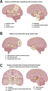
Brain regions associated with empathy. A) People instructed to look at interpersonal scenes and empathize with a specific person in an emotionally-charged vs. neutral situation showed greater activation in premotor cortex, thalamus, primary motor cortex, and primary somatosensory cortex. Participants simultaneously reported feeling similar emotions to the “other” person (Nummenmaa et al., 2008). B) Evidence for “group” emotions point to activation of the right posterior superior temporal sulcus and the left temporal pole, regions that are similarly activated when individuals feel pride in themselves (Takahashi et al., 2008). C) Other investigations into “group” feelings have shown that the striatum, substantia nigra, ventral tegmental area, insula, hippocampus, and amygdala have an increase in activation when sports fans watch emotionally-charged clips of their favorite teams (Duarte et al., 2017).
Fig. 3.
Fig. 3.
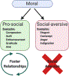
Schematic illustration of one of the ways social feelings can be broken down. Feelings of affiliation greatly depend on the individual’s perception of the other’s feelings. Generally, these can be grouped as having a negative or positive valence, and being self- or other-oriented. Additionally, morality is a large component of shared feelings, which can be grouped widely into pro-social or social-aversive. Examples of pro-social affiliative emotions include compassion, guilt, embarrassment, gratitude, and awe, and serve to build & foster relationships. Examples of social-aversive affiliative emotions include disgust, contempt, anger, and indignation, which often lead to social aversion or a break-down of potential relationships.
Fig. 4.
Fig. 4.
The social behavior neural network (Adapted from Smith et al., 2019a,b): Green boxes represent cortico-striatal regions; red box represents midbrain region; blue boxes represent hypothalamic regions.
Fig. 5.
Fig. 5.
Importance of oxytocin in pair bonding and maternal feelings. Oxytocin (OT) has been identified as an essential neurochemical in the formation of social attachment. Brain regions dense in OT and OT receptors (among other neuropeptides and monoamines) include the pre-optic anterior hypothalamic area, the septal region, and closely associated basal forebrain structures. Damage to this system interrupts naturally occurring monogamous pair-bonds in prairie voles, and formation of mother-child attachments.
Fig. 6.
Fig. 6.
The neurobiology of response to infant stimuli. Most of the literature investigating the neurobiology of mother-child attachment involves mothers watching scenes or videos of themselves with their children, or videos of their own babies or strange babies in various emotional states (e.g., happy, distressed, neutral). A) When mothers watched videos of themselves interacting with their own children, fMRI research shows increased activation in the dorsal anterior cingulate cortex, fusiform gyrus, cuneus, inferior parietal lobule, supplementary motor area, and nucleus accumbens. B) When mothers watched videos of their children, they generated greater activation in the amygdala and hypothalamus when their child was distressed as opposed to happy. C) Some research has investigated “high sensitivity” and” low sensitivity” mothers based on plasma oxytocin levels immediately following mother-child play. When shown their own child and a stranger’s child in neutral, happy or sad states, high sensitivity mothers displayed increased activation of the right superior temporal gyrus when their own child was happy compared to neutral. This was not seen in low-response mothers.
Fig. 7.
Fig. 7.
Parental affective neuroscience and response to infant stress. Parent-child relationships – like any relationship – are influenced by outside factors, such as previous childhood poverty experienced by the parents. A) Response to a distressed child shows sex-specific brain activation in parents who had experienced childhood poverty. Specifically, women show increased activity in the posterior insula, striatum, calcarine sulcus, hippocampus, and fusiform gyrus, whereas men show decreased activation in these regions in response to infant cries. These neurobiological changes were associated with self-reported feelings of annoyance and reduced desire to approach infants in both men and women. B) Intervention, such as training programs for promoting maternal empathy and learning stress reduction skills (called “Mom Power”), was shown to increase activity in typical child-focused, social brain areas like the precuneus, subgenual anterior cingulate cortex, and amygdala-temporal pole functional connectivity. This training and altered brain activity was accompanied by decreased annoyance and stress felt by mothers.
Fig. 8.
Fig. 8.
Regions associated with empathy, as informed by lesion and fMRI studies. Feelings of guilt and compassion are strongly associated with typical functioning of the subgenual/septal region and ventromedial frontal cortex. Longer-term emotions, such as processing long-term consequences and conceptualizing quality of social behavior, activate the frontopolar cortex and the right superior anterior temporal lobe.
Fig. 9.
Fig. 9.
Interpersonal stress and peripheral physiological responses. The physiological response to interpersonal stress has been investigated using a design where people were asked to “judge” another while imagining them in the judged-person’s position and rate their social feelings and “other” feelings. A) Social feelings assessed included i) submissiveness, ii) fear of losing social approval, iii) shame, iv) guilt, and v) embarrassment. B) Judges’ ratings of the other person’s feelings of submissiveness and fear of losing social approval predicted a larger increase in the judged person’s cortisol levels. C) Judges ratings of the other person’s feelings of embarrassment predicted a decrease in T-lymphocyte numbers.
Fig. 10.
Fig. 10.
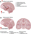
Neurobiology of interpersonal stress. Participants in these studies were asked to discuss their positive and negative qualities on video; they were then placed into a scanner, where they received “social feedback” from another person watching their video, which indicated adjectives such as serious or shallow. A) Greater release of the cytokine IL-6 was seen following trials, regardless of feedback type. Increased activity in the medial prefrontal cortex, posterior cingulate cortex, and hippocampus was observed when subjects reported increased momentary feelings of rejection, as well as increased activity in the amygdala and functional connectivity between the amygdala and dorsomedial prefrontal cortex. B) Another study examined responses to negative, positive, and neutral feedback given publicly or privately. This research led to the identification of the “Embarrassment Pathway,” which was most affected by negative feedback. This includes regions for processing the feedback, such as the dorsal anterior insula, processing the publicity, such as the medial prefrontal cortex and precuneus, and connectivity of these regions involved in affect processing, such as the amygdala and ventral anterior insula. These were also associated with increased pupil dilation.
Fig. 11.
Fig. 11.
Brain activity during emotional communication. A) When an individual perceives another person’s emotional behavior, the feelings elicited in the observer can be characterized as “shared” or “accompanying.” Accompanying feelings include pleasant feelings of confidence if the communication was successful (i.e. the feeling that one person correctly understood the other person’s feelings), feelings elicited when one partner regards the other partners emotional behavior as appropriate or not, induction of one’s own emotions regarding the other’s response, and assessing confidence in if the person understands the other’s feelings. B) Use of pseudo-hyperscanning allows researchers to l examine brain activity of a “sender” and a “perceiver” of emotional signals that can be temporally aligned. When a person is asked to communicate emotions via facial expressions, their communication partner shows similar neural activation. This is more pronounced between romantic partners than between strangers. The more similar the activation, the more shared feelings are reported (Anders et al., 2020b). C) Strangers viewing facial expressions of a sender show an increase in ventral striatum and medial orbitofrontal cortex activity that is correlated with the perceiver’s confidence in having correctly understood the sender’s emotion and predicts changes in interpersonal attraction (Anders et al., 2016). D) When people were asked to look at images of another person exhibiting publicly inappropriate behavior (a situation associated with self-reported feelings of Fremdscham or vicarious embarrassment caused by another’s inappropriate behavior), greater activations in the left insula and anterior cingulate cortex occurred, and decreased activation in the ventral striatum was observed. In such emerging studies there is no overt communication of feelings or emotions (perceivers inferred the targets’ feeling and emotional states from their actions in context or not at all), and the degree to which perceivers shared the targets’ feelings are not specifically measured. Future studies, though, may develop more robust paradigms to address these issues.
Fig. 12.
Fig. 12.
Neural networks of mental functions related to social feelings as revealed by the Neurosynth database. Left: Maps extract regions, marked in red, where activation occurs more consistently for studies that mention the term than for studies that do not. Right: Conjunction analysis indicating regions where neural networks overlap for two or more mental functions. Results are shown separately for the five “hot” and four “cold” mental functions (orange or blue color, respectively), and for all nine mental functions together (spectrum colors). Number of regionally overlapping functions is color coded as shown on respective scale. ACC anterior cingulate cortex, FMC frontomedian cortex, L left, OFC orbitofrontal cortex, PC precuneus, R right, SA subcallosal area, TP temporal pole, TPJ temporoparietal junction. Results for conjunction analyses are additionally illustrated in respective movies in the Supplement.

Similar articles

Cited by

References

    1. Adkins-Regan E, 2009. Neuroendocrinology of social behavior. ILAR J. 50, 5–14. - PubMed
    1. Adolphs R, Tranel D, Damasio H, Damasio AR, 1995. Fear and the human amygdala. J. Neurosci 15, 5879–5891. - PMC - PubMed
    1. Ainsworth MD, Bell SM, 1970. Attachment, exploration, and separation: illustrated by the behavior of one-year-olds in a strange situation. Child Dev. 41, 49–67. - PubMed
    1. Albers HE, 2012. The regulation of social recognition, social communication and aggression: vasopressin in the social behavior neural network. Horm. Behav 61, 283–292. - PubMed
    1. Albers HE, 2015. Species, sex and individual differences in the vasotocin/vasopressin system: relationship to neurochemical signaling in the social behavior neural network. Front. Neuroendocrinol 36, 49–71. - PMC - PubMed

Publication types