

Ventralis intermedius nucleus anatomical variability assessment by MRI structural connectivity
- PMID: 34089871
- PMCID: PMC8960999
- DOI: 10.1016/j.neuroimage.2021.118231
Ventralis intermedius nucleus anatomical variability assessment by MRI structural connectivity
Abstract
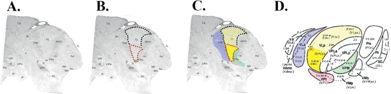
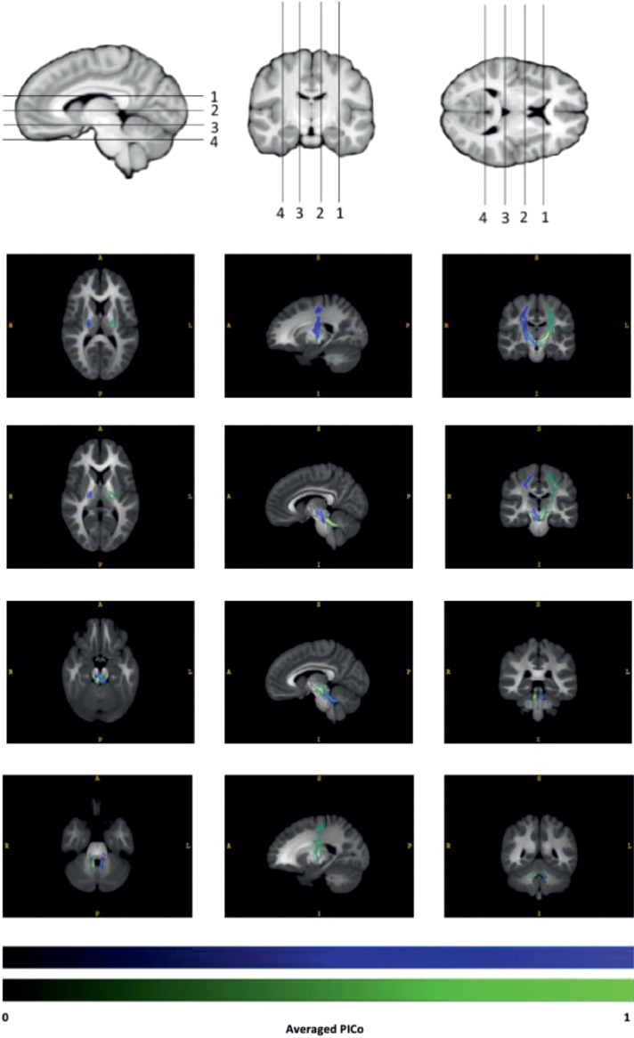
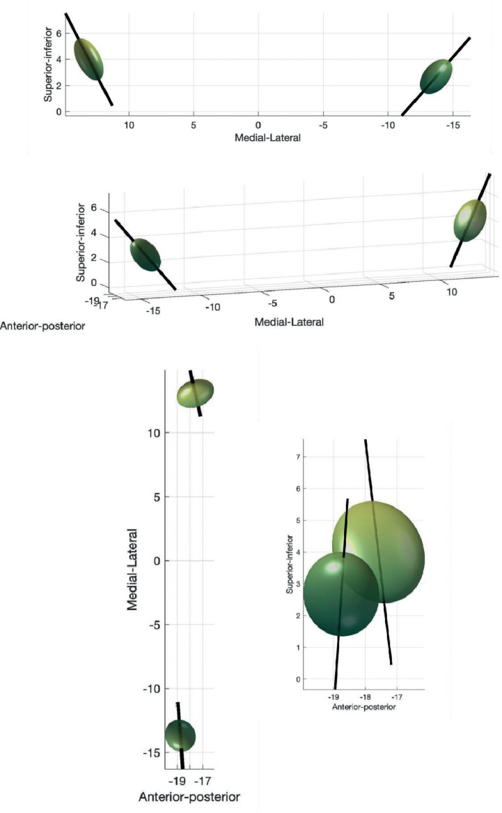
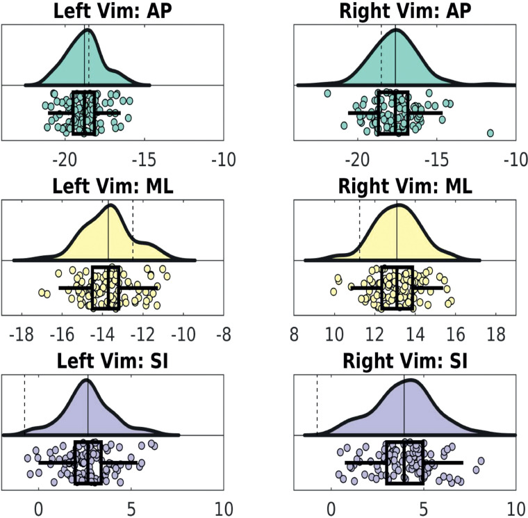
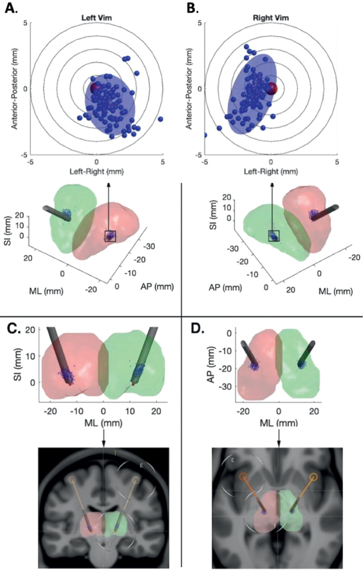
The ventralis intermedius nucleus (Vim) is centrally placed in the dentato-thalamo-cortical pathway (DTCp) and is a key surgical target in the treatment of severe medically refractory tremor. It is not visible on conventional MRI sequences; consequently, stereotactic targeting currently relies on atlas-based coordinates. This fails to capture individual anatomical variability, which may lead to poor long-term clinical efficacy. Probabilistic tractography, combined with known anatomical connectivity, enables localisation of thalamic nuclei at an individual subject level. There are, however, a number of confounds associated with this technique that may influence results. Here we focused on an established method, using probabilistic tractography to reconstruct the DTCp, to identify the connectivity-defined Vim (cd-Vim) in vivo. Using 100 healthy individuals from the Human Connectome Project, our aim was to quantify cd-Vim variability across this population, measure the discrepancy with atlas-defined Vim (ad-Vim), and assess the influence of potential methodological confounds. We found no significant effect of any of the confounds. The mean cd-Vim coordinate was located within 1.88 mm (left) and 2.12 mm (right) of the average midpoint and 3.98 mm (left) and 5.41 mm (right) from the ad-Vim coordinates. cd-Vim location was more variable on the right, which reflects hemispheric asymmetries in the probabilistic DTC reconstructed. The method was reproducible, with no significant cd-Vim location differences in a separate test-retest cohort. The superior cerebellar peduncle was identified as a potential source of artificial variance. This work demonstrates significant individual anatomical variability of the cd-Vim that atlas-based coordinate targeting fails to capture. This variability was not related to any methodological confound tested. Lateralisation of cerebellar functions, such as speech, may contribute to the observed asymmetry. Tractography-based methods seem sensitive to individual anatomical variability that is missed by conventional neurosurgical targeting; these findings may form the basis for translational tools to improve efficacy and reduce side-effects of thalamic surgery for tremor.
Keywords: Connectivity; Functional neurosurgery; Individualized targeting; Probabilistic tractography; Tremor.
Copyright © 2021. Published by Elsevier Inc.
Conflict of interest statement
Declaration of Competing Interest The authors declare no conflicts of interest, financial or otherwise.
Figures
Similar articles
-
Connectivity derived thalamic segmentation in deep brain stimulation for tremor.Neuroimage Clin. 2018 Jan 28;18:130-142. doi: 10.1016/j.nicl.2018.01.008. eCollection 2018. Neuroimage Clin. 2018. PMID: 29387530 Free PMC article.
-
Contrasting connectivity of the ventralis intermedius and ventralis oralis posterior nuclei of the motor thalamus demonstrated by probabilistic tractography.Neurosurgery. 2012 Jan;70(1):162-9; discussion 169. doi: 10.1227/NEU.0b013e3182262c9a. Neurosurgery. 2012. PMID: 22158304 Review.
-
Microelectrode recording findings within the tractography-defined ventral intermediate nucleus.J Neurosurg. 2017 May;126(5):1669-1675. doi: 10.3171/2016.3.JNS151992. Epub 2016 Jul 22. J Neurosurg. 2017. PMID: 27447439
-
Structural and functional connectivity of the nondecussating dentato-rubro-thalamic tract.Neuroimage. 2018 Aug 1;176:364-371. doi: 10.1016/j.neuroimage.2018.04.074. Epub 2018 May 4. Neuroimage. 2018. PMID: 29733955 Free PMC article.
-
The role of tractography in the localisation of the Vim nucleus of the thalamus and the dentatorubrothalamic tract for the treatment of tremor.Neurologia (Engl Ed). 2022 Oct;37(8):691-699. doi: 10.1016/j.nrleng.2019.09.008. Epub 2021 Sep 23. Neurologia (Engl Ed). 2022. PMID: 34563477 Review.
Cited by
-
When the central integrator disintegrates: A review of the role of the thalamus in cognition and dementia.Alzheimers Dement. 2024 Mar;20(3):2209-2222. doi: 10.1002/alz.13563. Epub 2023 Dec 2. Alzheimers Dement. 2024. PMID: 38041861 Free PMC article. Review.
-
Dynamic functional changes upon thalamotomy in essential tremor depend on baseline brain morphometry.Sci Rep. 2024 Jan 31;14(1):2605. doi: 10.1038/s41598-024-52410-y. Sci Rep. 2024. PMID: 38297028 Free PMC article.
-
FAT1-weighted MRI-guided focused ultrasound thalamotomy for essential tremor.BMJ Neurol Open. 2025 Jul 28;7(2):e001104. doi: 10.1136/bmjno-2025-001104. eCollection 2025. BMJ Neurol Open. 2025. PMID: 40734993 Free PMC article.
-
Deep brain stimulation for Parkinson's disease.J Intern Med. 2022 Nov;292(5):764-778. doi: 10.1111/joim.13541. Epub 2022 Jul 13. J Intern Med. 2022. PMID: 35798568 Free PMC article. Review.
-
Automatic planning of MR-guided transcranial focused ultrasound treatment for essential tremor.Front Neuroimaging. 2023 Oct 26;2:1272061. doi: 10.3389/fnimg.2023.1272061. eCollection 2023. Front Neuroimaging. 2023. PMID: 37953746 Free PMC article.
References
-
- Abhinav K., Yeh F.-C., Pathak S., Suski V., Lacomis D., Friedlander R.M., Fernandez-Miranda J.C. Advanced diffusion MRI fiber tracking in neurosurgical and neurodegenerative disorders and neuroanatomical studies: a review. Biochim. Biophys. Acta Mol. Basis Dis. 2014;1842:2286–2297. doi: 10.1016/j.bbadis.2014.08.002. - DOI - PubMed
-
- Akram H., Dayal V., Mahlknecht P., Georgiev D., Hyam J., Foltynie T., Limousin P., De Vita E., Jahanshahi M., Ashburner J., Behrens T., Hariz M., Zrinzo L. Connectivity derived thalamic segmentation in deep brain stimulation for tremor. NeuroImage Clin. 2018 doi: 10.1016/j.nicl.2018.01.008. - DOI - PMC - PubMed
-
- Alho Review of Printed and Electronic Stereotactic Atlases of the Human Brain. IntechOpe. 2011
Publication types
MeSH terms
Grants and funding
LinkOut - more resources
Full Text Sources
Miscellaneous
