

Use of post-mortem chest computed tomography in Covid-19 pneumonia
- PMID: 34090259
- PMCID: PMC8154189
- DOI: 10.1016/j.forsciint.2021.110851
Use of post-mortem chest computed tomography in Covid-19 pneumonia
Abstract
Background and aim: COVID-19 is an extremely challenging disease, both from a clinical and forensic point of view, and performing autopsies of COVID-19 deceased requires adequately equipped sectorial rooms and exposes health professionals to the risk of contagion. Among one of the categories that are most affected by SARS-Cov-2 infection are the elderly residents. Despite the need for prompt diagnoses, which are essential to implement all isolation measures necessary to contain the infection spread, deceased subjects in long-term care facilities are still are often diagnosed post-mortem. In this context, our study focuses on the use of post-mortem computed tomography for the diagnosis of COVID-19 infection, in conjunction with post-mortem swabs. The aim of this study was to assess the usefulness of post-mortem whole CT-scanning in identifying COVID-19 pneumonia as a cause of death, by comparing chest CT-findings of confirmed COVID-19 fatalities to control cases.
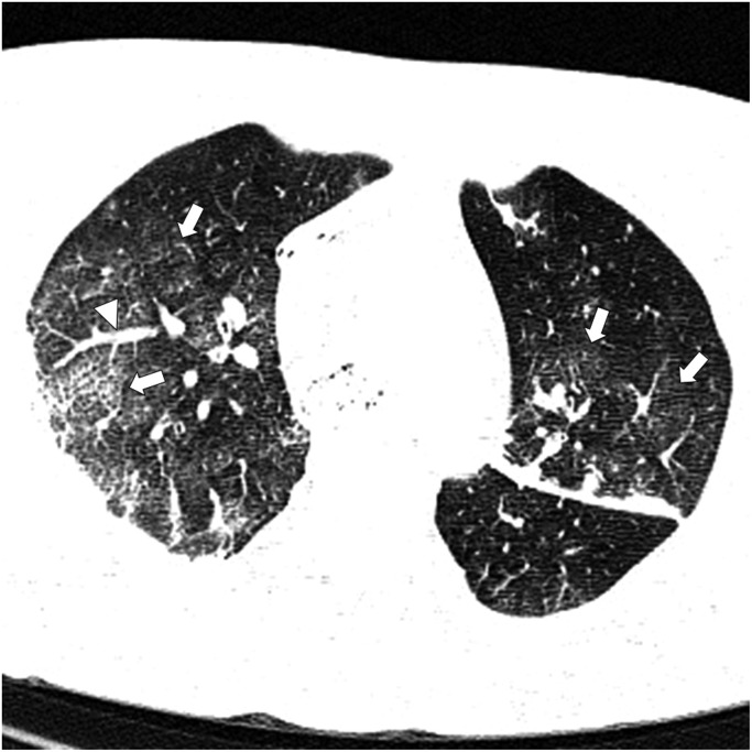
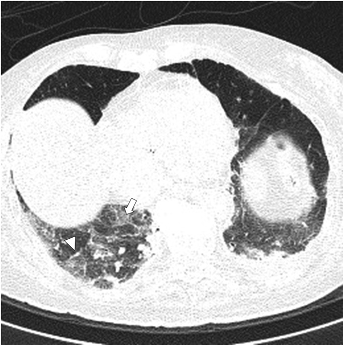
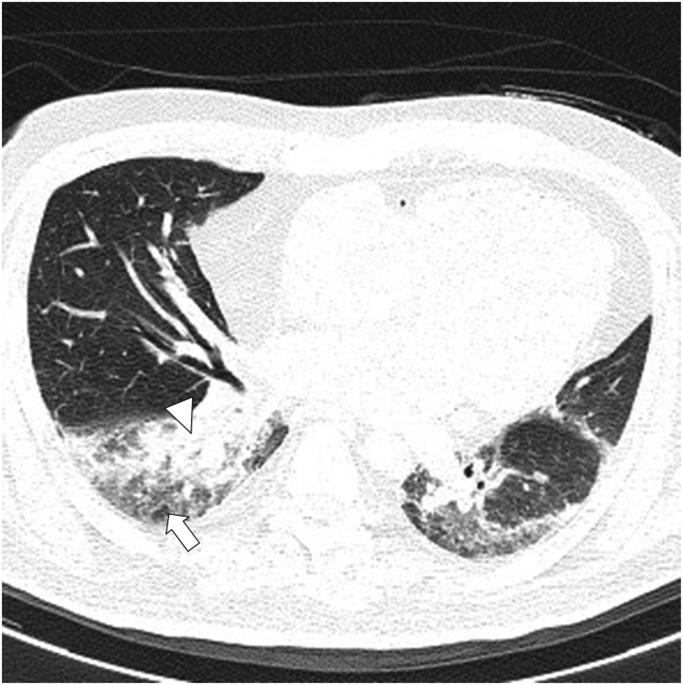
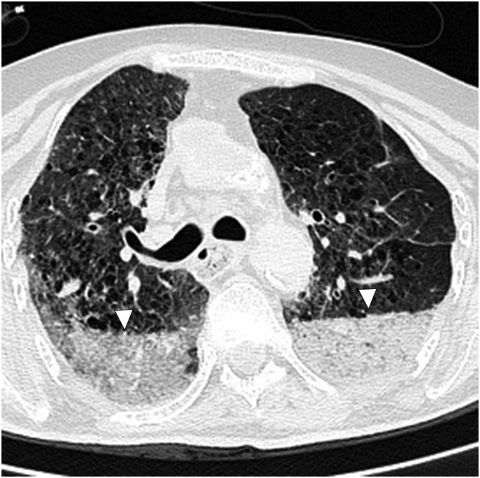
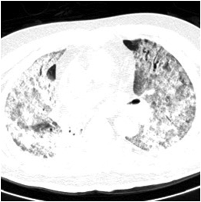
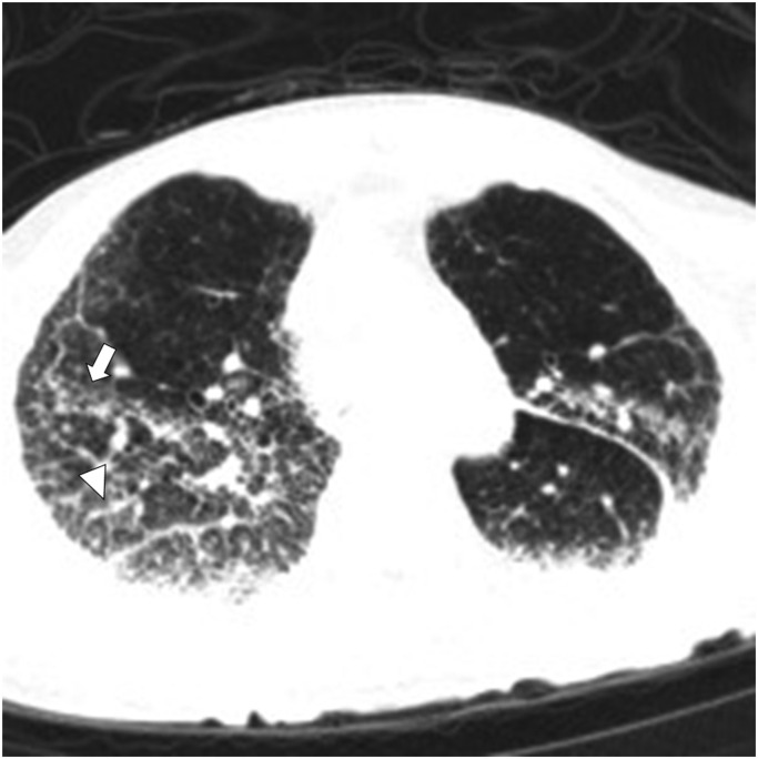
Materials and methods: The study included 24 deceased subjects: 13 subjects coming from long-term care facility and 11 subjects died at home. Whole body CT scans were performed within 48 h from death in all subjects to evaluate the presence and distribution of pulmonary abnormalities typical of COVID-19-pneumonia, including: ground-glass opacities (GGO), consolidation, and pleural effusion to confirm the post-mortem diagnosis.
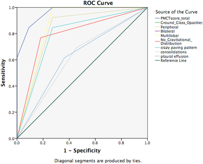
Results: Whole-body CT scans was feasible and allowed a complete diagnosis in all subjects. In 9 (69%) of the 13 cases from long-term care facility the cause of death was severe COVID 19 pneumonia, while GGO were present in 100% of the study population.
Conclusion: In the context of rapidly escalating COVID-19 outbreaks, given that laboratory tests for the novel coronavirus is time-consuming and can be falsely negative, the post-mortem CT can be considered as a reliable and safe modality to confirm COVID-19 pneumonia. This is especially true for specific postmortem chest CT-findings that are rather characteristic of COVID-19 fatalities.
Keywords: Autopsy; COVID-19; Coronavirus; Pneumonia; Post mortem changes; Post mortem computed tomography.
Crown Copyright © 2021. Published by Elsevier B.V. All rights reserved.
Conflict of interest statement
Declarations of interest None.
Figures

Similar articles
-
Complete post-mortem data in a fatal case of COVID-19: clinical, radiological and pathological correlations.Int J Legal Med. 2020 Nov;134(6):2209-2214. doi: 10.1007/s00414-020-02390-1. Epub 2020 Aug 6. Int J Legal Med. 2020. PMID: 32767018 Free PMC article.
-
COVID-19 pneumonia: CT findings of 122 patients and differentiation from influenza pneumonia.Eur Radiol. 2020 Oct;30(10):5463-5469. doi: 10.1007/s00330-020-06928-0. Epub 2020 May 12. Eur Radiol. 2020. PMID: 32399710 Free PMC article.
-
COVID-19 lungs in post-mortem computed tomography.Rechtsmedizin (Berl). 2021;31(2):145-147. doi: 10.1007/s00194-021-00462-z. Epub 2021 Feb 15. Rechtsmedizin (Berl). 2021. PMID: 33612977 Free PMC article.
-
Potentials of post-mortem CT investigations during SARS-COV-2 pandemic: a narrative review.Radiol Med. 2022 Apr;127(4):383-390. doi: 10.1007/s11547-022-01457-w. Epub 2022 Feb 28. Radiol Med. 2022. PMID: 35226246 Free PMC article. Review.
-
Similarities and Differences of Early Pulmonary CT Features of Pneumonia Caused by SARS-CoV-2, SARS-CoV and MERS-CoV: Comparison Based on a Systemic Review.Chin Med Sci J. 2020 Sep 30;35(3):254-261. doi: 10.24920/003727. Chin Med Sci J. 2020. PMID: 32972503 Free PMC article.
Cited by
-
Regarding "Post-mortem CT lung findings at a medicolegal institute in SARS-CoV-2 RT-PCR positive cases with autopsy correlation".Forensic Sci Med Pathol. 2022 Mar;18(1):114-115. doi: 10.1007/s12024-021-00430-9. Epub 2021 Nov 1. Forensic Sci Med Pathol. 2022. PMID: 34724159 Free PMC article. No abstract available.
-
Postmortem Chest Computed Tomography in Fatal COVID-19: A Valuable Diagnostic Tool for Minimally Invasive Autopsy.Clinics (Sao Paulo). 2021 Dec 8;76:e3551. doi: 10.6061/clinics/2021/e3551. eCollection 2021. Clinics (Sao Paulo). 2021. PMID: 34909914 Free PMC article. No abstract available.
-
Korean radiographers' awareness, experiences, and education needs in forensic medicine and forensic radiology.Heliyon. 2024 May 31;10(11):e32219. doi: 10.1016/j.heliyon.2024.e32219. eCollection 2024 Jun 15. Heliyon. 2024. PMID: 38873674 Free PMC article.
-
Dying "from" or "with" COVID-19 during the Pandemic: Medico-Legal Issues According to a Population Perspective.Int J Environ Res Public Health. 2021 Aug 22;18(16):8851. doi: 10.3390/ijerph18168851. Int J Environ Res Public Health. 2021. PMID: 34444600 Free PMC article.
-
Postmortem chest computed tomography in COVID-19: A minimally invasive autopsy method.Eur J Radiol Open. 2024 Jan 13;12:100546. doi: 10.1016/j.ejro.2024.100546. eCollection 2024 Jun. Eur J Radiol Open. 2024. PMID: 38293283 Free PMC article.
References
-
- World Health Organization, Coronavirus disease (COVID-2019) situation reports. Accessed April 3, 2020.
MeSH terms
LinkOut - more resources
Full Text Sources
Medical
Miscellaneous
