. 2021 Jun 5;20(1):84.
doi: 10.1186/s12943-021-01380-0.

CHEK1 and circCHEK1_246aa evoke chromosomal instability and induce bone lesion formation in multiple myeloma

Affiliations

CHEK1 and circCHEK1_246aa evoke chromosomal instability and induce bone lesion formation in multiple myeloma

Chunyan Gu et al. Mol Cancer. .

Abstract

Background: Multiple myeloma (MM) is still incurable and characterized by clonal expansion of plasma cells in the bone marrow (BM). Therefore, effective therapeutic interventions must target both myeloma cells and the BM niche.

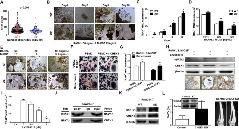
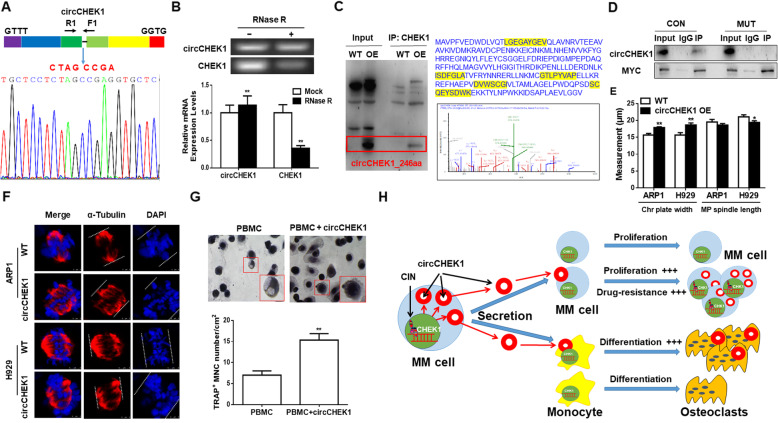
Methods: Cell proliferation, drug resistance, and chromosomal instability (CIN) induced by CHEK1 were confirmed by Giemsa staining, exon sequencing, immunofluorescence and xenograft model in vivo. Bone lesion was evaluated by Tartrate-resistant acid phosphatase (TRAP) staining. The existence of circCHEK1_246aa was evaluated by qPCR, Sanger sequencing and Mass Spectrometer.

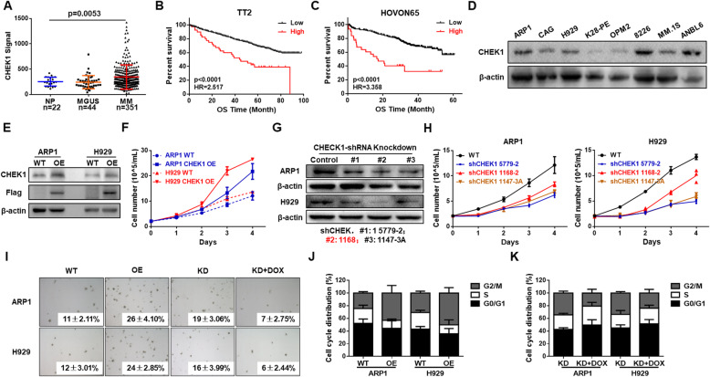
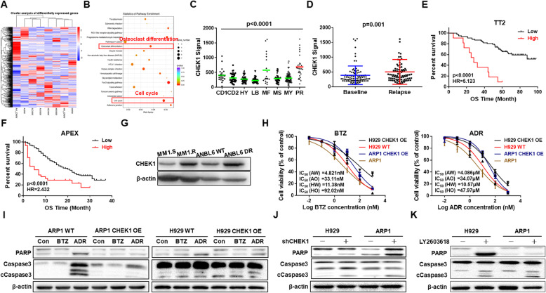
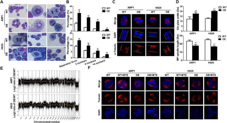
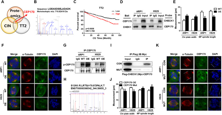
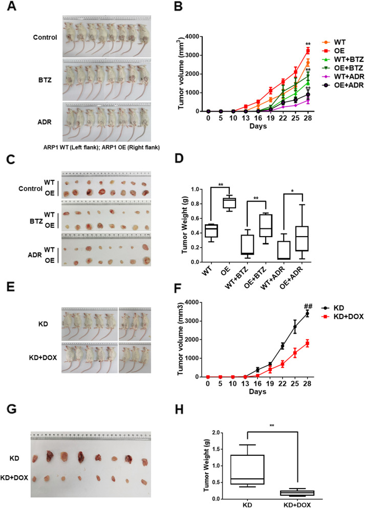
Results: We demonstrated that CHEK1 expression was significantly increased in human MM samples relative to normal plasma cells, and that in MM patients, high CHEK1 expression was associated with poor outcomes. Increased CHEK1 expression induced MM cellular proliferation and evoked drug-resistance in vitro and in vivo. CHEK1-mediated increases in cell proliferation and drug resistance were due in part to CHEK1-induced CIN. CHEK1 activated CIN, partly by phosphorylating CEP170. Interestingly, CHEK1 promoted osteoclast differentiation by upregulating NFATc1 expression. Intriguingly, we discovered that MM cells expressed circCHEK1_246aa, a circular CHEK1 RNA, which encoded and was translated to the CHEK1 kinase catalytic center. Transfection of circCHEK1_246aa increased MM CIN and osteoclast differentiation similarly to CHEK1 overexpression, suggesting that MM cells could secrete circCHEK1_246aa in the BM niche to increase the invasive potential of MM cells and promote osteoclast differentiation.

Conclusions: Our findings suggest that targeting the enzymatic catalytic center encoded by CHEK1 mRNA and circCHEK1_246aa is a promising therapeutic modality to target both MM cells and BM niche.

Keywords: CHEK1; Chromosomal instability; Drug resistance; Multiple myeloma; Proliferation; circCHEK1_246aa.


Conflict of interest statement

The authors declare they have no competing interests.

Figures

Fig. 1
Fig. 1
Elevated CHEK1 expression is associated with poor outcomes in MM patients and promotes MM cell proliferation in vitro. A CHEK1 mRNA levels were significantly increased in MM samples. The signal level of CHEK1 is shown on the y-axis. Patients were designated as being healthy donors with normal bone marrow plasma cells (NP, n = 22), monoclonal gammopathy of undetermined significance (MGUS, n = 44), or multiple myeloma (MM, n = 351), and are sorted on the x-axis. B Increased CHEK1 mRNA expression was associated with poor overall survival (OS) in MM patients from the TT2 patient cohort. C Increased CHEK1 mRNA expression was associated with poor OS in MM patients from the HOVON65 cohort. D Western blot analysis revealed that CHEK1 was endogenously expressed in the specified MM cell lines. E Validation of CHEK1 overexpression (OE) in CHEK1-OE ARP1 and H929 cells relative to vehicle-transfected control cells (WT). F Four-day cell growth curve, as detected by trypan blue staining and counting of WT, CHEK1-OE ARP1, and H929 cells. G Confirmation of CHEK1 protein knockdown (KD) in ARP1 and H929 cells after transfection with three independent CHEK1-targeting shRNAs. H Four-day cell growth curve in WT, CHEK1-KD ARP1, and H929 cells. I Images of representative soft agar plates, revealing increased clonogenic growth of CHEK1-OE cells and decreased clonogenic growth in CHEK1-KD cells relative to WT. J Cell cycle analysis revealed that the proportion of G2/M phase cells significantly increased in CHEK1-OE cells relative to WT. K Cell cycle analysis revealed that the proportion of G2/M phase cells significantly decreased in CHEK1-KD cells
Fig. 2
Fig. 2
CHEK1 is a marker for high-risk MM and induces drug resistance. A Heatmap of RNA-seq data showing significantly differentiated genes before and after doxycycline-induced CHEK1 OE. B Pathway enrichment analysis of RNA-seq data revealed enrichment of two pathways, which were related to cell cycle regulation and osteoclast differentiation. C Box plot representing CHEK1 expression in eight MM risk subgroups from the TT2 patient cohort. D In paired patient MM samples collected at first diagnosis and relapse, CHEK1 mRNA expression was increased in the relapsed samples relative to the corresponding samples from first diagnosis. E–F Increased CHEK1 expression was correlated with decreased OS in relapsed patients from the (E) TT2 and (F) APEX cohorts. G Western blotting confirmed that CHEK1 protein levels were significantly increased in MM1.R (dexamethasone-resistant) and ANBL6 DR (Bortezomib-resistant) cells. H Effects of Bortezomib and Adriamycin on the cell viability of H929 and ARP1 cells with or without CHEK1 OE. I Western blots demonstrated that CHEK1 OE induced resistance to Adriamycin and Bortezomib in ARP1 and H929 cells, as indicated by cleavage of the apoptotic regulators PARP and Caspase 3. J–K Pro-apoptotic effects of (J) CHEK1 shRNA silencing and the (K) CHEK1 selective inhibitor LY2603618 in H929 and ARP1 cells, as demonstrated by increased cleavage of PARP and Caspase 3
Fig. 3
Fig. 3
CHEK1 evokes chromosomal instability (CIN) in MM. A–B Giemsa staining revealed that CHEK1 OE increased the separation error rate and number of multi-nuclear cells in (A) ARP1 and (B) H929 cells. C–D Increased chromosomal plate width and decreased mitotic bipolar spindle length in CHEK1-OE ARP1 and H929 cells relative to WT, as demonstrated by immunofluorescent (IF) staining for α-tubulin and DAPI. E A comparative genomic hybridization (CGH) array revealed significant gains and losses of multiple chromosomal segments in CHEK1-OE ARP1 and H929 cells relative to WT. F In WT and CHEK1-OE cells treated with vehicle or Borbezomib, chromosomal plate width was highest and mitotic spindle length lowest in the Borbezomib-treated CHEK1-OE group
Fig. 4
Fig. 4
CHEK1 promotes CIN through CEP170 activation in MM. A–B Centrosomal Protein 170 (CEP170) was selected among candidate genes of the CIN-related gene list and genes associated with poor outcome in the TT2 MM patient cohort. C Increased CEP170 expression was associated with decreased OS in the TT2 patient cohort. D A Co-IP assay revealed that CHEK1 directly interacted with CEP170 in CHEK1-OE ARP1 and H929 cells. E–F CEP170 OE significantly increased chromosomal plate width and decreased mitotic bipolar spindle length in ARP1 and H929 cells. G A Co-IP assay confirmed that CHEK1 physically interacted with and phosphorylated CEP170 in CHEK1-OE cells compared with WT cells, as detected by total anti-phospho-serine antibody. H Mass spectrometry (MS) was used to determine the CHEK1 phosphorylation site of CEP170, Ser1260. I A Myc-tagged CEP170 Ser1260Ala mutant, containing a defective CHEK1 phosphorylation site, exhibited dramatically decreased interaction with flag-tagged CHEK1, as demonstrated by Co-IP followed by western blotting. J–K OE of mutated CEP170 Ser1260Ala decreased chromosomal plate width and increased mitotic bipolar spindle length in (J) ARP1 and (K) H929 cells
Fig. 5
Fig. 5
CHEK1 induces macrophage osteoclast by upregulating NFATc1 expression. A Magnetic resonance imaging (MRI) revealed that increased CHEK1 expression was positively correlated with bone lesion formation in TT2 cohort MM patients. B–C TRAP staining revealed that Chek1 OE promoted osteoclast differentiation in RAW 264.7 mouse macrophages co-treated with RANKL (50 ng/mL) and M-CSF (15 ng/mL) in a time-dependent manner. D–E TRAP staining confirmed that Chek1 OE prompted osteoclast differentiation in RAW 264.7 cells treated with varying doses of RANKL and M-CSF in a manner dependent on RANKL and M-CSF dosages. F–G TRAP staining revealed that human primary peripheral blood mononuclear cells (PBMCs) transfected with human CHEK1 cDNA developed significant more osteoclasts than non-transfected control cells. H–I Western blotting and TRAP staining confirmed that the CHEK1 inhibitor LY2603618 decreased NFATc1 expression and suppressed osteoclast differentiation in RAW 264.7 cells. J Co-IP revealed that CHEK1 interacted with NFATc1 in RAW 264.7 cells. K Western blotting confirmed that the expression of NFATc1 was increased in Chek1-OE RAW264.7 cells relative to WT cells. L CHEK1 knockdown prevented myeloma-associated bone loss in 5TMM3VT model. Micro-CT analysis of 5TMM3VT-involved tibia bone performed at 4 weeks confirmed the presence of osteolytic lesions and demonstrated decreased trabecular bone volume (BV/TV) compared with CHEK1 gene knockdown
Fig. 6
Fig. 6
MM cells secrete circCHEK1_246aa circular RNA to induce MM CIN and promote osteoclast differentiation in the bone marrow microenvironment. A The number of exons and exact circCHEK1 sequences produced from CHEK1 were validated by Sanger sequencing. The blue arrow represents the “head-to-tail” splicing sites of circCHEK1. B mRNA levels of circCHEK1 and linear CHEK1 ± RNase R were determined by RT-PCR and qRT-PCR. C After pull-down using a CHEK1 antibody, protein samples at the expected size were excised and subjected to mass spectrometry (MS) analysis, and specific peptides from circCHEK1_246aa were identified. D A Co-IP assay revealed that circCHEK1_246aa more robustly interacted with native CEP170 than mutated CEP170. E–F circCHEK1 OE increased chromosomal plate width and decreased mitotic bipolar spindle length in ARP1 and H929 cells. G TRAP staining revealed that circCHEK1-OE human primary PBMCs developed into significantly more osteoclasts relative to vehicle-transfected control cells. H Graphic illustrating that CHEK1 and circCHEK1_246aa promote multiple myeloma malignancy by evoking CIN and bone lesion formation
Fig. 7
Fig. 7
CHEK1 promotes MM growth in vivo and is a potential therapeutic target. A Photographic images of xenograft-bearing mice from each group were taken at day 28. B Time course of tumor growth in NOD-SCID mice treated with vehicle, BTZ, or ADR. C Photographic images of xenografts from NOD-SCID mice of the specified groups on day 28. D Mean tumor weights in the six experimental groups at day 28 after implantation of the specified MM cells. E Photographic images of xenograft-bearing mice from the KD and KD + DOX groups were collected at day 28. F Time course of tumor growth in the NOD-SCID mice of the specified groups. G Xenografts from the NOD-SCID mice of the specified groups were collected at day 28. H Mean tumor weights in the specified two experimental groups at day 28 after implantation of MM cells

Similar articles

Cited by

References

    1. Bianchi G, Anderson KC. Understanding biology to tackle the disease: multiple myeloma from bench to bedside, and back. CA Cancer J Clin. 2014;64(6):422–444. doi: 10.3322/caac.21252. - DOI - PubMed
    1. Pawlyn C, Morgan GJ. Evolutionary biology of high-risk multiple myeloma. Nat Rev Cancer. 2017;17(9):543–556. doi: 10.1038/nrc.2017.63. - DOI - PubMed
    1. Gu C, Lu T, Wang W, Shao M, Wei R, Guo M, et al. RFWD2 induces cellular proliferation and selective proteasome inhibitor resistance by mediating P27 ubiquitination in multiple myeloma. Leukemia. 2020. 10.1038/s41375-020-01033-z. - PubMed
    1. Lohr JG, Stojanov P, Carter SL, Cruz-Gordillo P, Lawrence MS, Auclair D, Sougnez C, Knoechel B, Gould J, Saksena G, Cibulskis K, McKenna A, Chapman MA, Straussman R, Levy J, Perkins LM, Keats JJ, Schumacher SE, Rosenberg M, Getz G, Golub TR, Anderson KC, Richardson P, Krishnan A, Lonial S, Kaufman J, Siegel DS, Vesole DH, Roy V, Rivera CE, Rajkumar SV, Kumar S, Fonseca R, Ahmann GJ, Bergsagel PL, Stewart AK, Hofmeister CC, Efebera YA, Jagannath S, Chari A, Trudel S, Reece D, Wolf J, Martin T, Zimmerman T, Rosenbaum C, Jakubowiak AJ, Lebovic D, Vij R, Stockerl-Goldstein K. Widespread genetic heterogeneity in multiple myeloma: implications for targeted therapy. Cancer Cell. 2014;25(1):91–101. doi: 10.1016/j.ccr.2013.12.015. - DOI - PMC - PubMed
    1. Gu C, Cheng H, Yang H, Bian Y, Wang Y, Zhang Y, Pisano M, Hu G, Yang Y. MK2 is a therapeutic target for high-risk multiple myeloma. Haematologica. 2018;1:1. doi: 10.3324/haematol.2017.182121. - DOI - PMC - PubMed

Publication types