

Sequence determinants of in cell condensate morphology, dynamics, and oligomerization as measured by number and brightness analysis
- PMID: 34090478
- PMCID: PMC8178893
- DOI: 10.1186/s12964-021-00744-9
Sequence determinants of in cell condensate morphology, dynamics, and oligomerization as measured by number and brightness analysis
Abstract
Background: Biomolecular condensates are non-stoichiometric assemblies that are characterized by their capacity to spatially concentrate biomolecules and play a key role in cellular organization. Proteins that drive the formation of biomolecular condensates frequently contain oligomerization domains and intrinsically disordered regions (IDRs), both of which can contribute multivalent interactions that drive higher-order assembly. Our understanding of the relative and temporal contribution of oligomerization domains and IDRs to the material properties of in vivo biomolecular condensates is limited. Similarly, the spatial and temporal dependence of protein oligomeric state inside condensates has been largely unexplored in vivo.
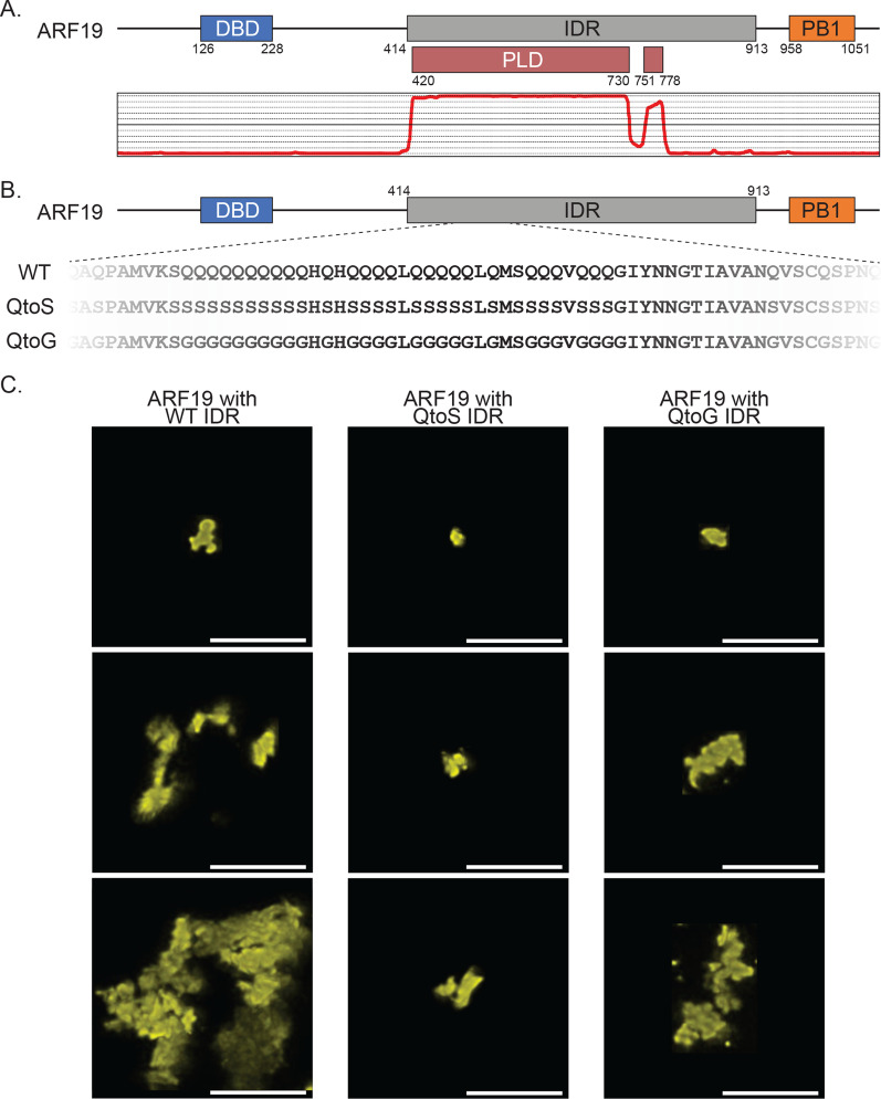
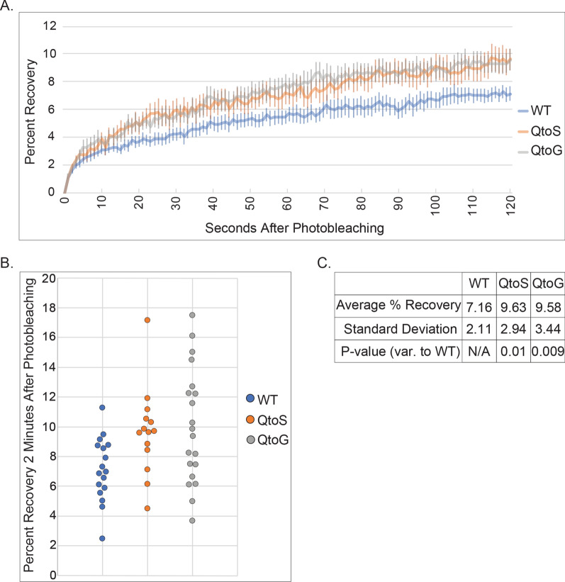
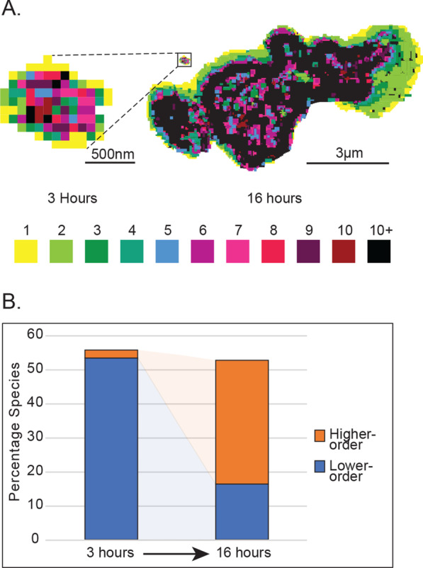
Methods: In this study, we combined quantitative microscopy with number and brightness analysis to investigate the aging, material properties, and protein oligomeric state of biomolecular condensates in vivo. Our work is focused on condensates formed by AUXIN RESPONSE FACTOR 19 (ARF19), a transcription factor integral to the auxin signaling pathway in plants. ARF19 contains a large central glutamine-rich IDR and a C-terminal Phox Bem1 (PB1) oligomerization domain and forms cytoplasmic condensates.
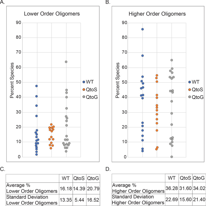
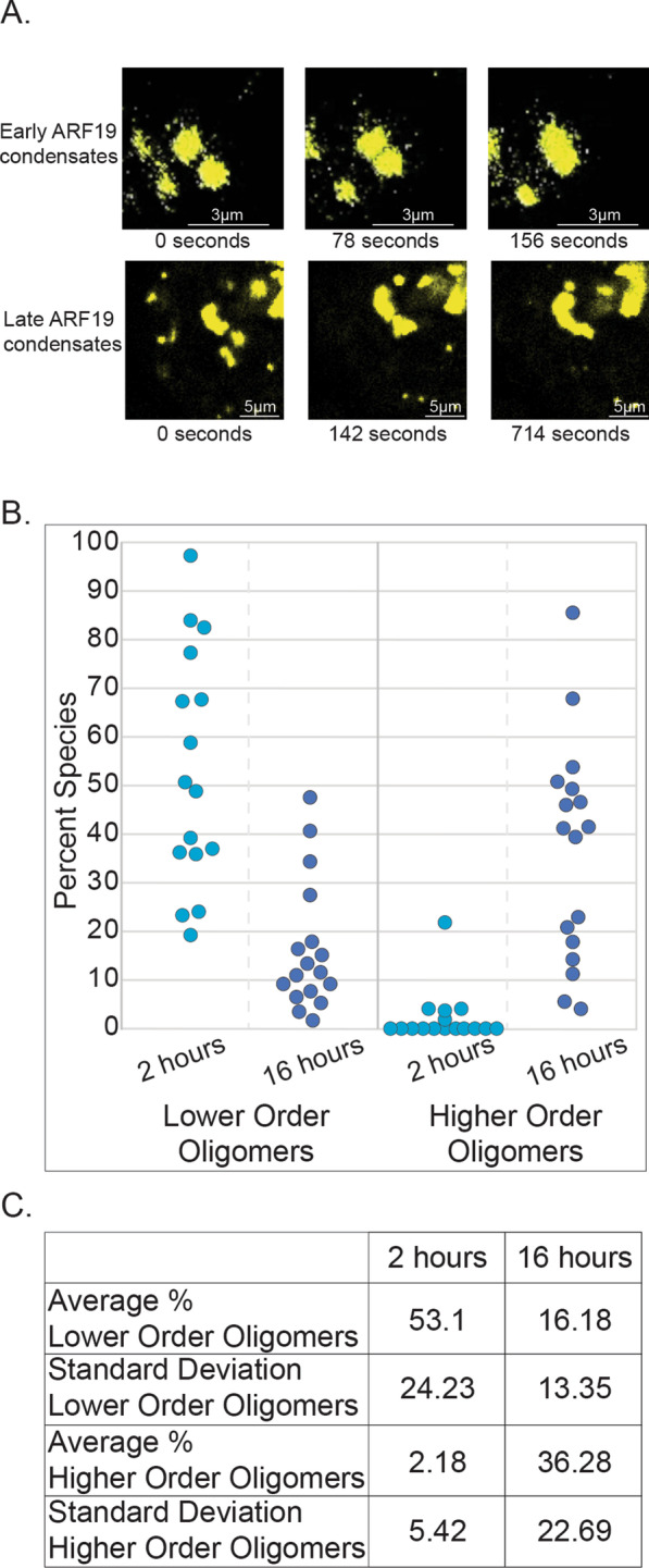
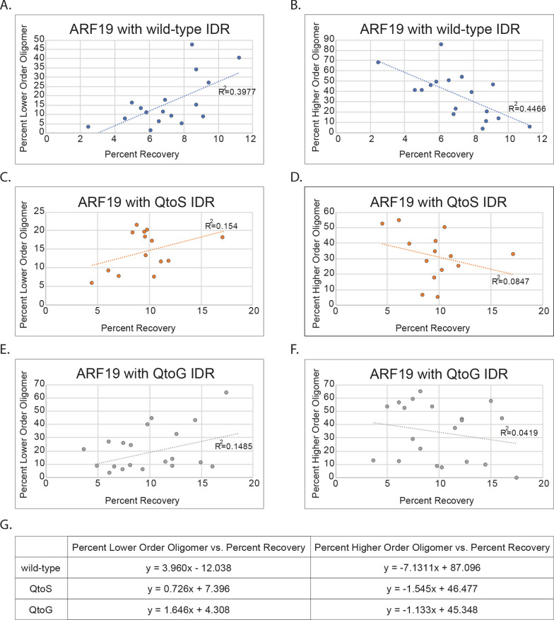
Results: Our results reveal that the IDR amino acid composition can influence the morphology and material properties of ARF19 condensates. In contrast the distribution of oligomeric species within condensates appears insensitive to the IDR composition. In addition, we identified a relationship between the abundance of higher- and lower-order oligomers within individual condensates and their apparent fluidity.
Conclusions: IDR amino acid composition affects condensate morphology and material properties. In ARF condensates, altering the amino acid composition of the IDR did not greatly affect the oligomeric state of proteins within the condensate. Video Abstract.
Keywords: Biomolecular condensates; Fluorescence microscopy; Fluorescence recovery after photobleaching; Intrinsically disordered regions; Number and brightness analysis.
Conflict of interest statement
A.S.H. is a scientific consultant with Dewpoint Therapeutics. R.J.E. and L.C.S. declare no competing interests.
Figures
Similar articles
-
Active transport enables protein condensation in cells.Sci Adv. 2025 May 23;11(21):eadv7875. doi: 10.1126/sciadv.adv7875. Epub 2025 May 23. Sci Adv. 2025. PMID: 40408482 Free PMC article.
-
Disorder-mediated interactions target proteins to specific condensates.Mol Cell. 2024 Sep 19;84(18):3497-3512.e9. doi: 10.1016/j.molcel.2024.08.017. Epub 2024 Sep 3. Mol Cell. 2024. PMID: 39232584
-
Intrinsically disordered regions and RNA binding domains contribute to protein enrichment in biomolecular condensates in Xenopus oocytes.Sci Rep. 2024 Nov 13;14(1):27890. doi: 10.1038/s41598-024-79409-9. Sci Rep. 2024. PMID: 39537752 Free PMC article.
-
Conformational Dynamics of Intrinsically Disordered Proteins Regulate Biomolecular Condensate Chemistry.Chem Rev. 2022 Mar 23;122(6):6719-6748. doi: 10.1021/acs.chemrev.1c00774. Epub 2022 Feb 18. Chem Rev. 2022. PMID: 35179885 Free PMC article. Review.
-
Current perspectives in drug targeting intrinsically disordered proteins and biomolecular condensates.BMC Biol. 2025 May 6;23(1):118. doi: 10.1186/s12915-025-02214-x. BMC Biol. 2025. PMID: 40325419 Free PMC article. Review.
Cited by
-
Phase Transitions of Associative Biomacromolecules.Chem Rev. 2023 Jul 26;123(14):8945-8987. doi: 10.1021/acs.chemrev.2c00814. Epub 2023 Mar 7. Chem Rev. 2023. PMID: 36881934 Free PMC article. Review.
-
FER-like iron deficiency-induced transcription factor (FIT) accumulates in nuclear condensates.J Cell Biol. 2024 Apr 1;223(4):e202311048. doi: 10.1083/jcb.202311048. Epub 2024 Feb 23. J Cell Biol. 2024. PMID: 38393070 Free PMC article.
-
Shedding light on iron nutrition: exploring intersections of transcription factor cascades in light and iron deficiency signaling.J Exp Bot. 2025 Feb 7;76(3):787-802. doi: 10.1093/jxb/erae324. J Exp Bot. 2025. PMID: 39115876 Free PMC article. Review.
-
Mechanosensitive ion channels MSL8, MSL9, and MSL10 have environmentally sensitive intrinsically disordered regions with distinct biophysical characteristics in vitro.Plant Direct. 2023 Aug 3;7(8):e515. doi: 10.1002/pld3.515. eCollection 2023 Aug. Plant Direct. 2023. PMID: 37547488 Free PMC article.
-
Active transport enables protein condensation in cells.Sci Adv. 2025 May 23;11(21):eadv7875. doi: 10.1126/sciadv.adv7875. Epub 2025 May 23. Sci Adv. 2025. PMID: 40408482 Free PMC article.
References
Publication types
MeSH terms
Substances
Grants and funding
LinkOut - more resources
Full Text Sources
Miscellaneous
