. 2021 Jun 5;12(1):322.
doi: 10.1186/s13287-021-02384-9.

Bone marrow mesenchymal stem cells and their derived exosomes resolve doxorubicin-induced chemobrain: critical role of their miRNA cargo

Affiliations

Bone marrow mesenchymal stem cells and their derived exosomes resolve doxorubicin-induced chemobrain: critical role of their miRNA cargo

Marwa O El-Derany et al. Stem Cell Res Ther. .

Abstract

Background: Doxorubicin (DOX), a widely used chemotherapeutic agent, can cause neurodegeneration in the brain, which leads to a condition known as chemobrain. In fact, chemobrain is a deteriorating condition which adversely affects the lives of cancer survivors. This study aimed to examine the potential therapeutic effects of bone marrow mesenchymal stem cells (BMSCs) and their derived exosomes (BMSCs-Exo) in DOX-induced chemobrain in rat models.

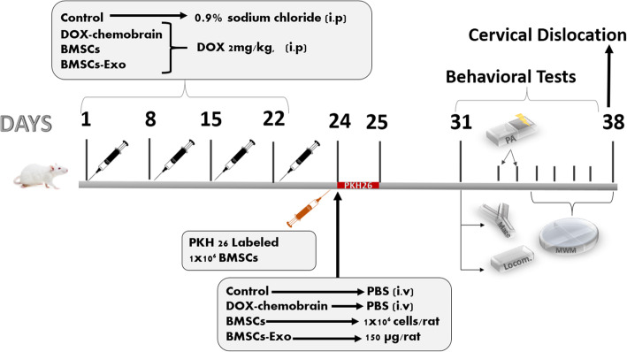
Methods: Chemobrain was induced by exposing rats to DOX (2 mg/kg, i.p) once weekly for 4 consecutive weeks. After 48 h of the last DOX dose, a subset of rats was supplied with either an intravenous injection of BMSCs (1 × 10⁶) or a single dose of 150 μg of BMSCs-Exo. Behavioral tests were conducted 7 days post injection. Rats were sacrificed after 14 days from BMSCs or BMSCs-Exo injection.

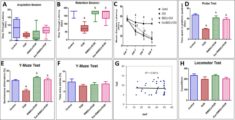
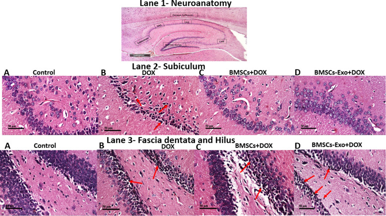
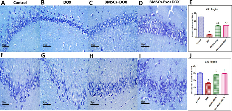
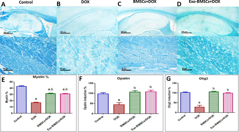
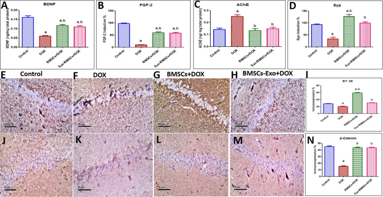
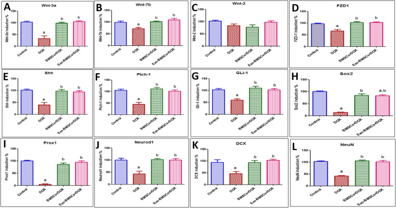
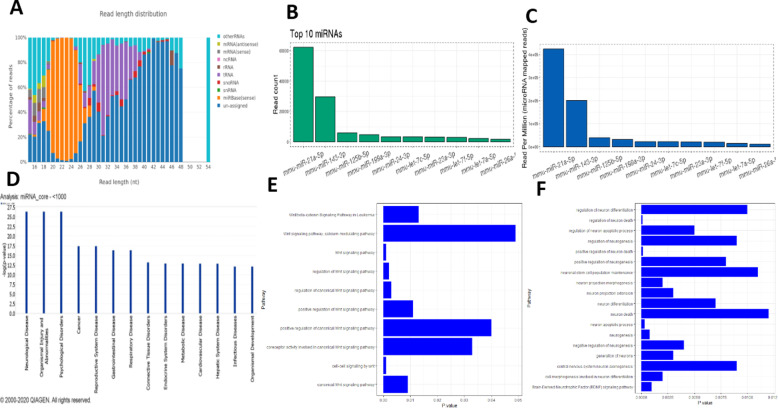
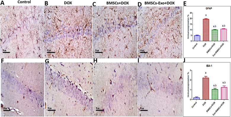
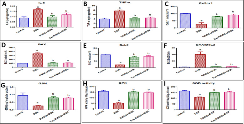
Results: BMSCs and BMSCs-Exo successfully restored DOX-induced cognitive and behavioral distortion. These actions were mediated via decreasing hippocampal neurodegeneration and neural demyelination through upregulating neural myelination factors (myelin%, Olig2, Opalin expression), neurotropic growth factors (BDNF, FGF-2), synaptic factors (synaptophysin), and fractalkine receptor expression (Cx3cr1). Halting neurodegeneration in DOX-induced chemobrain was achieved through epigenetic induction of key factors in Wnt/β-catenin and hedgehog signaling pathways mediated primarily by the most abundant secreted exosomal miRNAs (miR-21-5p, miR-125b-5p, miR-199a-3p, miR-24-3p, let-7a-5p). Moreover, BMSCs and BMSCs-Exo significantly abrogate the inflammatory state (IL-6, TNF-α), apoptotic state (BAX/Bcl2), astrocyte, and microglia activation (GFAP, IBA-1) in DOX-induced chemobrain with a significant increase in the antioxidant mediators (GSH, GPx, SOD activity).

Conclusions: BMSCs and their derived exosomes offer neuroprotection against DOX-induced chemobrain via genetic and epigenetic abrogation of hippocampal neurodegeneration through modulating Wnt/β-catenin and hedgehog signaling pathways and through reducing inflammatory, apoptotic, and oxidative stress state. Proposed mechanisms of the protective effects of bone marrow stem cells (BMSCs) and their exosomes (BMSCs-Exo) in doxorubicin (DOX)-induced chemobrain. Blue arrows: induce. Red arrows: inhibit.

Keywords: BMSCs; Chemobrain; Exosomes; Signaling pathway; miRNAs.


Conflict of interest statement

The authors have read the journal’s policy on disclosure of potential conflicts of interest and they all declare no personal or financial conflict of interest.

Figures

Fig. 1
Fig. 1
An illustration of the study design showing the timeline of the induction of chemobrain by doxorubicin (DOX) and the treatment with bone marrow stem cells (BMSCs) or their exosomes (BMSCs-Exo), and behavioral test schedule. Rats were randomly assigned into four groups (n = 20/group) and treated for 4 weeks as follows: The first group served as the control group and received intra-peritoneal (i.p) injection of 0.9% sodium chloride given once weekly for 4 consecutive weeks. Forty-eight hours later, a single intravenous injection of 150 μL of particle-free PBS was given. The second group served as DOX-treated group and received DOX hydrochloride dissolved in 0.9% sodium chloride and given once weekly in a dose of 2 mg/kg, i.p. for 4 consecutive weeks. Forty-eight hours later, a single intravenous injection of 150 μL of particle-free PBS was given. The third group received DOX once weekly (2 mg/kg, i.p.) for 4 consecutive weeks followed by a single intravenous injections of (1 × 10⁶) BMSCs per rat that was given 48 h after the last DOX dose. The fourth group was the BMSCs-Exo-treated group and received DOX once weekly (2 mg/kg, i.p.) for 4 consecutive weeks followed by a single dose of 150 μg of exosomal proteins per rat that was given 48 h after last DOX dose. Four rats were treated by labeled PKH26 Red Fluorescent Cell Linker BMSCs, and after 24 h, rats were then anesthetized with ketamine (100 mg/kg, i.p.) and sacrificed by cervical dislocation the whole brains were excised. Behavioral testing started 7 days after BMSCs and BMSCs-Exo administration. After 14 days from BMSCs and BMSCs-Exo injection, the four groups were then anesthetized with ketamine (100 mg/kg, i.p.) and sacrificed by cervical dislocation, the whole brains were excised, and hippocampi were dissected and weighed
Fig. 2
Fig. 2
Characterization, differentiation, and migration of bone marrow stem cells (BMSCs). A A representative image showing fibroblastic morphology of BMSCs (× 200). B Flow cytometry curve showing a control, left side: gating of unstained cells at FITC, right side: gating of unstained cells at PE. C Flow cytometry curve showing the expression of CD105 in BMSCs. D Flow cytometry curve showing the expression of CD73 in BMSCs. E Flow cytometry curve showing the expression of CD34 in BMSCs. F Flow cytometry curve showing the expression of CD14 expression in BMSCs. G Flow cytometry curve showing the expression of CD63 in exosome produced by BMSCs. H Control BMSCs cultured in LDMEM supplemented with 10% bovine serum, 100 U/mL penicillin, and 100 μg/mL streptomycin, left side: stained with Oil Red and right side: stained with Alizarin Red (× 200). I BMSCs stained with Oil Red showing adipogenic differentiation of BMSCs (× 200). J BMSCs stained with Alizarin Red showing osteogenic differentiation of BMSCs (× 200). K Transmission electron microscope image of exosome produced by BMSCs showing vesicular structure (arrow). L Localization of PKH26-labeled BMSCs in the hippocampal regions of the brain tissues of rats (× 200) (n = 4)
Fig. 3
Fig. 3
Bone marrow stem cell (BMSC) and their exosome (BMSCs-Exo) treatment reverse behavioral changes induced by doxorubicin (DOX) in rats. A, B Box plots representing results from step-through passive avoidance behavioral test. C A scatter plot representing results from Morris water maze test. D A bar chart representing results from the probe test. E A bar chart representing Y-maze % of alternation (SAP). F A bar chart representing Y-maze total arm entries (TAE). G A scatter plot representing the correlation between SAP and TAE and H a bar chart representing results from the locomotor activity test. For A and B, data are presented as medians (25th, 75th percentile) and statistical analysis was carried out using Kruskal–Wallis non-parametric test followed by Dunn’s test (n = 8). For CH, data are presented as mean ± SEM (n = 8). For C, statistical analysis was performed using repeated two-way ANOVA followed by the Bonferroni post-tests as post hoc test. For D–F and H, statistical analysis was performed using one-way ANOVA followed by Tukey’s test as post hoc test (n = 8). a Significantly different from the control group at p < 0.05. b Significantly different from DOX group at p < 0.05. For G, statistical analysis was performed using Pearson’s correlation
Fig. 4
Fig. 4
Histopathological analysis of rat brain sections using H&E staining. Lane (1) (× 100): Neuroanatomy of brain areas being investigated to show brain structures in the representative micrographs. Lane (2) (× 400): A Control group showed normal histological structure of the neurons in the subiculum. B DOX-induced chemobrain group showed nuclear pyknosis and degeneration observed in diffuse manner all over the neurons in the subiculum (arrow). C BMSC-treated DOX group showed normal subiculum histological features. D BMSCs-Exo treated DOX group showed no histological alterations in the subiculum regions. Lane (3) (× 400): A the control group showed normal histological structure of the neurons in the fascia dentata and hilus of the hippocampus. B DOX-induced chemobrain group showed nuclear pyknosis and degeneration observed in fascia dentata and hilus (arrow) of the hippocampus. C BMSC-treated DOX group showed few nuclear pyknosis and degeneration in fascia dentata and hilus of the hippocampus (arrow). D BMSCs-Exo-treated DOX group showed nuclear pyknosis and degeneration was shown in some neurons in fascia dentata and hilus of the hippocampal region (arrow) (n = 4)
Fig. 5
Fig. 5
Bone marrow stem cell (BMSC) and their exosome (BMSCs-Exo) treatment reverse neural damage induced by doxorubicin (DOX) in rats. Representative images of toluidine blue-stained brain tissue sections (× 400) of A the control group showing normal histological structure showing intact neurons in CA1 region, B DOX-induced chemobrain group showing decrease in intact neuron in CA1 region, C BMSC treatment group showing significant restoration in intact neurons in CA1 region, and D BMSCs-Exo treatment group showing significant restoration in intact neurons in CA1 region. E A bar chart reflecting the quantitative image analysis for intact neurons in CA1 region expressed intact cell number per field. Representative images of toluidine blue-stained brain tissue sections (× 400) of F control group showing normal histological structure showing intact neurons in CA3 region, G DOX-induced chemobrain group showing decrease in intact neuron in CA3 region, H BMSC treatment group showing significant restoration in intact neurons in CA3 region, and I BMSCs-Exo treatment group showing significant restoration in intact neurons in CA3 region. J A bar chart reflecting the quantitative image analysis for intact neurons in CA3 region expressed intact cell number per field. For E and J, statistical analysis was performed using one-way ANOVA followed by Tukey’s test as post hoc test (n = 4). Data are presented as mean ± SEM. a Significantly different from the control group at p < 0.05. b Significantly different from DOX group at p < 0.05
Fig. 6
Fig. 6
Bone marrow stem cell (BMSC) and their exosome (BMSCs-Exo) treatment prevent neural demyelination induced by doxorubicin (DOX) in rats. Representative images of Luxol fast blue-stained sections (× 100 and × 400) of A the control group showing positive stained myelinated nerve fibers in the corpus callosum region, B DOX-induced chemobrain group showing decrease in positively stained myelinated nerve fibers in the corpus callosum region, C BMSC treatment group showing restoration of positively stained myelinated nerve fibers in the corpus callosum region, and D BMSCs-Exo treatment group showing restoration of positively stained myelinated nerve fibers in the corpus callosum region. E A bar chart reflecting the quantitative image analysis for positive stained myelinated nerve fibers in the corpus callosum region expressed as area percent. F A bar chart representing the % of mRNA expression induction of Opalin in different rat groups. G A bar chart representing the % of mRNA expression induction of Olig-2 in different rat groups. For E, F, and G, statistical analysis was performed using one-way ANOVA followed by Tukey’s test as post hoc test (n = 6). Data are presented as mean ± SEM. a Significantly different from the control group at p < 0.05. b Significantly different from the DOX group at p < 0.05
Fig. 7
Fig. 7
Bone marrow stem cell (BMSC) and their exosome (BMSCs-Exo) treatment restore hippocampal neurotropic and synaptic growth factor, functional neurotransmitters, and hippocampal β-catenin expression in doxorubicin (DOX)-induced chemobrain in rats. A A bar chart representing the protein expression induction of BDNF in different rat groups. B A bar chart representing the % of mRNA expression induction of FGF-2 in different rat groups. C A bar chart representing the quantification of acetylcholinesterase (AChE) in different rat groups. D A bar chart representing the % of mRNA expression induction of Syp in different rat groups. Representative image reflecting immunohistochemical staining of SY 38 in E control group showing moderate expression of SY 38 in hippocampal sections, F DOX-induced chemobrain group showing minimal expression of SY 38, G group treated with BMSCs showing marked expression of SY 38, and H group treated with BMSCs-Exo showing moderate expression of SY 38. I A bar plot reflecting the quantitative image analysis for SY 38 immunohistochemical staining expressed as area percent. Representative image reflecting immunohistochemical staining of β-catenin in J the control group showing marked expression of β-catenin in hippocampal region, K DOX-induced chemobrain group showing minimal expression of β-catenin in hippocampal region, L BMSC-treated group showing marked expression of β-catenin, and M BMSCs-Exo-treated group showing marked expression of β-catenin. N A bar plot reflecting quantitative image analysis for β-catenin immunohistochemical staining expressed as area percent. Data in AD (n = 6), I, N (n = 4) are presented as mean ± SEM. Statistical analysis was performed using one-way ANOVA followed by Tukey’s post hoc test. a Significantly different from the control group at p < 0.05. b Significantly different from the DOX group at p < 0.05
Fig. 8
Fig. 8
Effects of bone marrow stem cell (BMSC) and their exosome (BMSCs-Exo) treatment on Wnt/β-catenin and hedgehog signaling genes and transcription factors in doxorubicin (DOX)-induced chemobrain in rats. A A bar chart representing the % of mRNA expression induction of Wnt-3a in different rat groups. B A bar chart representing the % of mRNA expression induction of Wnt-7b in different rat groups. C A bar chart representing the % of mRNA expression induction of Wnt-2 in different rat groups. D A bar chart representing the % of mRNA expression induction of FZD1 in different rat groups. E A bar chart representing the % of mRNA expression induction of sonic hedgehog (shh) in different rat groups. F A bar chart representing the % of mRNA expression induction of patched-1 (Ptch-1) in different rat groups. G A bar chart representing the % of mRNA expression induction of the transcription factor Gli-1 in different rat groups. H A bar chart representing the % of mRNA expression induction of Sox2 in different rat groups. I A bar chart representing the % of mRNA expression induction of Prox1 in different rat groups. J A bar chart representing the % of mRNA expression induction of NeuroD1 in different rat groups. K A bar chart representing the % of mRNA expression induction of DCX in different rat groups. L A bar chart representing the % of mRNA expression induction of NeuN in different rat groups. Data are presented as mean ± SEM. Statistical analysis was performed using one-way ANOVA followed by Tukey’s post hoc test (n = 6). a Significantly different from the control group at p < 0.05. b Significantly different from the DOX group at p < 0.05
Fig. 9
Fig. 9
Analysis of the miRNA cargo of exosomes secreted by bone marrow stem cells (BMSC-Exo). A A bar plot representing the distribution of sizes of the sequenced RNA molecules in BMSC-Exo. B A bar plot reflecting the counts of the top 10 most abundant miRNA in BMSC-Exo. C A bar plot reflecting the normalized counts of the top 10 most abundant miRNA in BMSC-Exo. D A bar plot reflecting the association significance between the discovered miRNA and disease as predicted by IPA analysis. E Association between miR-21-5p and Wnt/β-catenin signaling based on interaction data from the miRPathDB (10.1093/nar/gkz1022). F Association between miR-21-5p and neural hallmarks based on interaction data from the miRPathDB (10.1093/nar/gkz1022)
Fig. 10
Fig. 10
Bone marrow stem cells (BMSCs) and their exosomes (BMSCs-Exo) halt astrocytes and microglia activation in doxorubicin (DOX)-induced chemobrain in rats. Representative image reflecting immunohistochemical staining of GFAP “a marker of astrocyte activation” in A the control group showing minimal expression of GFAP in hippocampal region, B DOX-induced chemobrain group showing high expression of GFAP in hippocampal region, C BMSC-treated group showing moderate expression of GFAP, and D BMSCs-Exo-treated group showing moderate expression of GFAP. E A bar plot reflecting quantitative image analysis for GFAP immunohistochemical staining expressed as area percent. Representative image reflecting immunohistochemical staining of IBA-1 “a marker of microglia activation” in F the control group showing minimal expression of IBA-1 in the hippocampal region, G DOX-induced chemobrain group showing high expression of IBA-1 in hippocampal region, H BMSC-treated group showing moderate expression of IBA-1, and I BMSCs-Exo-treated group showing moderate expression of IBA-1. J A bar plot reflecting quantitative image analysis for IBA-1 immunohistochemical staining expressed as area percent. Data are presented as mean ± SEM (n = 4). Statistical analysis was performed using one-way ANOVA followed by Tukey’s test as post hoc test. a Significantly different from the control group at p < 0.05. b Significantly different from the DOX group at p < 0.05
Fig. 11
Fig. 11
Effects of bone marrow stem cell (BMSC) and their exosome (BMSCs-Exo) treatment on inflammatory, apoptotic, and oxidative stress markers in doxorubicin (DOX)-induced chemobrain in rats. A A bar chart representing the protein expression of interleukin (IL)-6 in different rat groups. B A bar chart representing the protein expression of tumor necrosis factor (TNF)-α in different rat groups. C A bar chart representing the % of mRNA expression induction of Cx3cr1 in different rat groups. D A bar chart representing the % of mRNA expression induction of BAX in different rat groups. E A bar chart representing the % of mRNA expression induction of Bcl-2 in different rat groups. F A bar chart representing BAX/Bcl2 ratio in different rat groups. G A bar chart representing the abundance of glutathione (GSH) in brains of different rat groups. H A bar chart representing the activity of glutathione peroxidase enzyme (GPx) in brains of different rat groups. I A bar chart representing the activity of superoxide dismutase enzyme (SOD) in brains of different rat groups. Data are presented as mean ± SEM. Statistical Analysis was performed using one-way ANOVA followed by Tukey’s post hoc test (n = 6). a Significantly different from the control group at p < 0.05. b Significantly different from the DOX group at p < 0.05

Similar articles

Cited by

References

    1. Dietrich J. Chemotherapy associated central nervous system damage. Adv Exp Med Biol. 2010;678:77–85. doi: 10.1007/978-1-4419-6306-2_11. - DOI - PubMed
    1. Aluise CD, Sultana R, Tangpong J, Vore M, St Clair D, Moscow JA, Butterfield DA. Chemo brain (chemo fog) as a potential side effect of doxorubicin administration: role of cytokine-induced, oxidative/nitrosative stress in cognitive dysfunction. Adv Exp Med Biol. 2010;678:147–156. doi: 10.1007/978-1-4419-6306-2_19. - DOI - PubMed
    1. Mauer ME, Bottomley A, Taphoorn MJB. Evaluating health-related quality of life and symptom burden in brain tumour patients: instruments for use in experimental trials and clinical practice. Curr Opin Neurol. 2008;21(6):745–753. doi: 10.1097/WCO.0b013e328315ef7d. - DOI - PubMed
    1. Wigmore P. The effect of systemic chemotherapy on neurogenesis, plasticity and memory. Curr Top Behav Neurosci. 2013;15:211–240. doi: 10.1007/7854_2012_235. - DOI - PubMed
    1. McLauchlan D, Robertson NP. Stem cells in the treatment of central nervous system disease. J Neurol. 2018;265(4):984–986. doi: 10.1007/s00415-018-8818-7. - DOI - PMC - PubMed