

Cardiac iron overload evaluation in thalassaemic patients using T2* magnetic resonance imaging following chelation therapy: a multicentre cross-sectional study
- PMID: 34090847
- PMCID: PMC9938451
- DOI: 10.1016/j.htct.2021.01.014
Cardiac iron overload evaluation in thalassaemic patients using T2* magnetic resonance imaging following chelation therapy: a multicentre cross-sectional study
Abstract
Introduction: Magnetic resonance imaging (MRI) T2* technique is used to assess iron overload in the heart, liver and pancreas of thalassaemic patients. Optimal iron chelation and expected tissue iron response rates remain under investigation. The objective of this study was to analyse serum ferritin and the iron concentration in the heart, liver and pancreas measured by MRI T2*/R2* during regular chelation therapy in a real-world cohort of patients with thalassemia.
Methods: We evaluated thalassaemic patients ≥ 7 years old undergoing chelation/transfusion therapy by MRI and assessed serum ferritin at baseline and follow-up from 2004-2011.
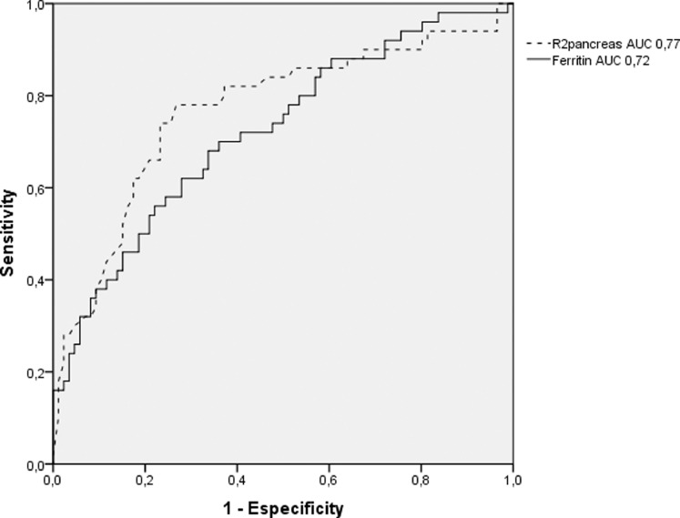
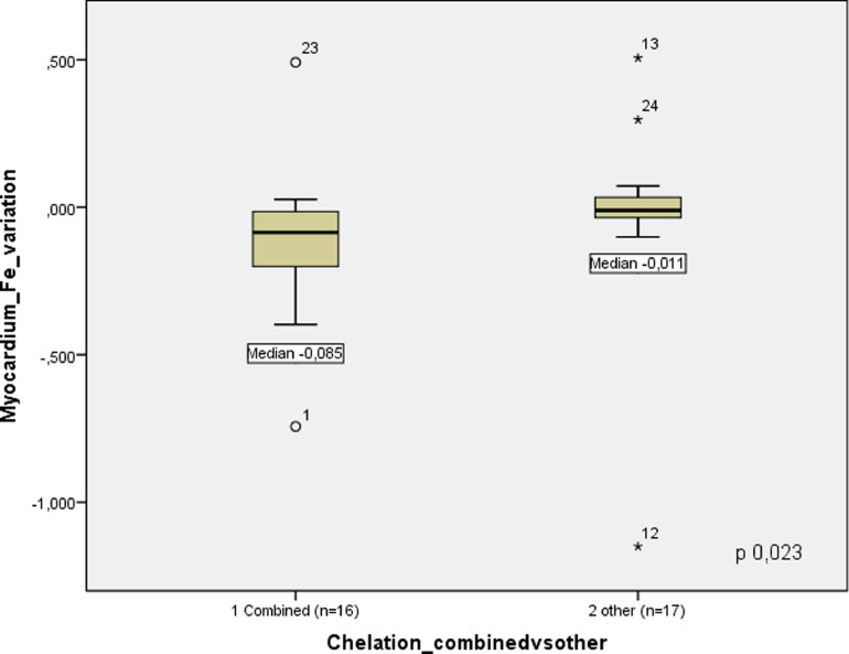
Results: We evaluated 136 patients, 92% major thalassaemic, with a median age of 18 years, and median baseline ferritin 2.033ng/ml (range: 59-14,123). Iron overload distribution was: liver (99%), pancreas (74%) and heart (36%). After a median of 1.2 years of follow-up, the iron overload in the myocardium reduced from 2,63 Fe mg/g to 2,05 (p 0.003). The optimal R2* pancreas cut-off was 148 Hertz, achieving 78% sensitivity and 73% specificity. However, when combining the R2* pancreas cut off ≤ 50 Hertz and a ferritin ≤ 1222 ng/ml, we could reach a negative predictive value (NPV) of 98% for cardiac siderosis. Only 28% were undergoing combined chelation at baseline assessment, which increased up to 50% on follow up evaluation.
Conclusions: Chelation therapy significantly reduced cardiac siderosis in thalassaemic patients. In patients with moderate/severe liver iron concentration undergoing chelation therapy, ferritin levels and myocardium iron improved earlier than the liver siderosis.
Keywords: Chelation therapy; Iron overload; Magnetic resonance imaging; Thalassemia.
Copyright © 2021. Published by Elsevier España, S.L.U.
Figures
References
-
- Betts M, Flight PA\, Paramore LC, Tian L, Milenković D, Sheth S. Systematic literature review of the burden of disease and treatment for transfusion-dependent β-thalassemia. Clin Ther. 2019;42(2) 322–337.e2. - PubMed
-
- Davis BA, Porter JB. Long-term outcome of continuous 24-hour deferoxamine infusion via indwelling intravenous catheters in high-risk beta-thalassemia. Blood. 2000;95(4):1229–1236. - PubMed
-
- Anderson LJ, Holden S, Davis B, Prescott E, Charrier CC, Bunce NH, et al. Cardiovascular T2-star (T2*) magnetic resonance for the early diagnosis of myocardial iron overload. Eur Heart J. 2001;22(23):2171–2179. - PubMed
-
- Fu C, Kannengiesser S, Cheng S, Shen J, Dong H, Yan F. Quantitative analysis of hepatic iron in patients suspected of coexisting iron overload and steatosis using multi-echo single-voxel magnetic resonance spectroscopy: Comparison with fat-saturated multi-echo gradient echo sequence. J Magn Reson Imaging. 2018 Jul;48(1):205–213. - PubMed
-
- Tanner MA, He T, Westwood MA, Firmin DN, Pennell DJ, Thalassemia International Federation Heart T2* Investigators Multi-center validation of the transferability of the magnetic resonance T2* technique for the quantification of tissue iron. Haematologica. 2006;91(10):1388–1391. - PubMed
LinkOut - more resources
Full Text Sources
