Review
. 2021 Aug:49:92-101.
doi: 10.1016/j.coviro.2021.05.002. Epub 2021 Jun 3.

Structural perspectives on HCV humoral immune evasion mechanisms

Affiliations
Review

Structural perspectives on HCV humoral immune evasion mechanisms

Madhumati Sevvana et al. Curr Opin Virol. 2021 Aug.

Abstract

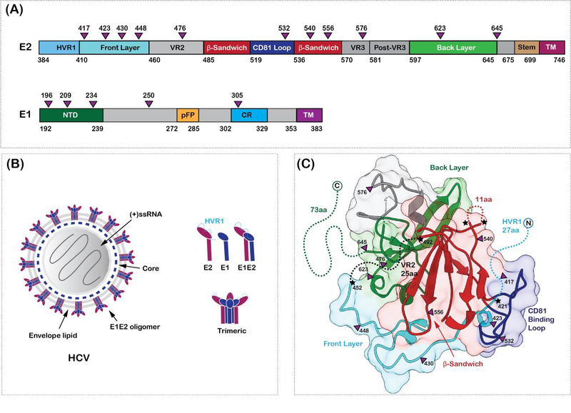
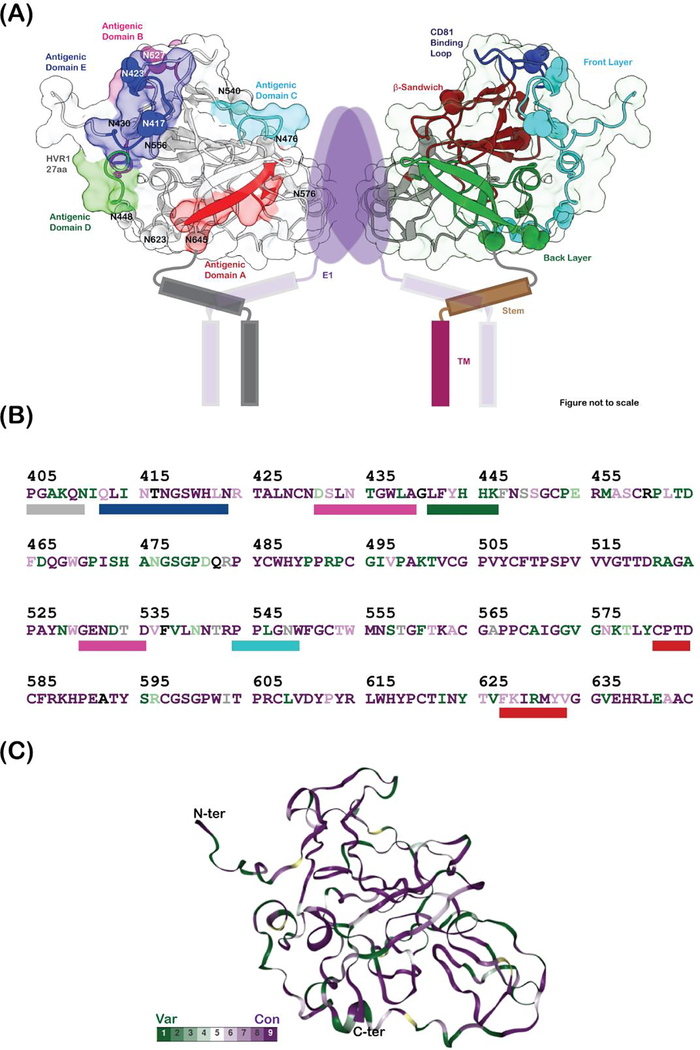
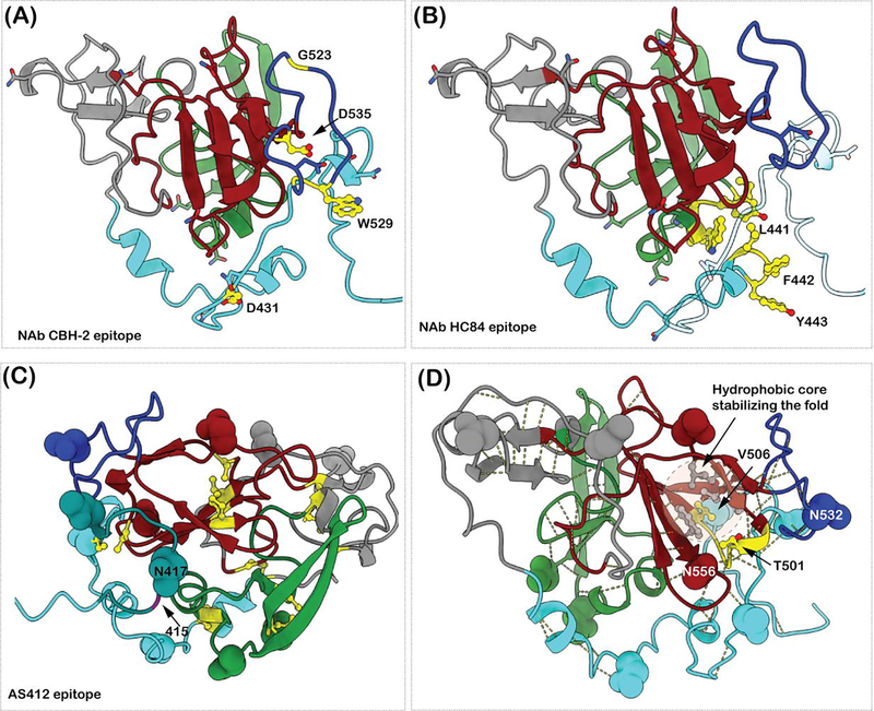
The molecular mechanisms of hepatitis C virus (HCV) persistence and pathogenesis are poorly understood. The design of an effective HCV vaccine is challenging despite a robust humoral immune response against closely related strains of HCV. This is primarily because of the huge genetic diversity of HCV and the molecular evolution of various virus escape mechanisms. These mechanisms are steered by the presence of a high mutational rate in HCV, structural plasticity of the immunodominant regions on the virion surface of diverse HCV genotypes, and constant amino acid substitutions on key structural components of HCV envelope glycoproteins. Here, we review the molecular basis of neutralizing antibody (nAb)-mediated immune response against diverse HCV variants, HCV-steered humoral immune evasion strategies and explore the essential structural elements to consider for designing a universal HCV vaccine. Structural perspectives on key escape pathways mediated by a point mutation within the epitope, allosteric modulation of the epitope by distant mutations and glycan shift on envelope glycoproteins will be highlighted (abstract graphic).


Conflict of interest statement

Declaration of interests

The authors declare that they have no known competing financial interests or personal relationships that could have appeared to influence the work reported in this paper.

Figures

Figure 1.
Figure 1.. HCV envelope glycoproteins.
(A) Domain organization of HCV envelope glycoproteins, E1 and E2. The glycosylation sites are shown as purple triangles in (A) and (C). The hypervariable region 1 (HVR1), variable regions 2 and 3 (HVR2 and HVR3), the front layer, the back layer, the β-sandwich domains, stem and the transmembrane (TM) regions are labelled on E2. The N-terminal domain (NTD), putative fusion peptide (pFP), the conserved region (CR) and the transmembrane regions are labelled on E1. (B) Hepatitis C virion architecture. E1 (dark blue) and E2 (dark purple) form heterodimers which in turn might arrange into trimers on the virus surface. The components of the virion are labelled. (C) Crystal structure of E2 core domain (PDB ID: 4MWF). Missing regions in the structure are shown as dotted lines and highlighted with a star. The coloring scheme is similar to (A)
Figure 2.
Figure 2.. The immunogenic regions of HCV E2.
(A) Cartoon representation of heterodimeric E1E2 proteins. The dimer of E1E2 heterodimers representation was prepared to simultaneously depict the antigenic regions and the secondary structure definitions. This does not represent the functional oligomeric state of the E1E2 heterodimers on the virus surface. Figure prepared from a homology model of E2 (residues 405–645) calculated using the program modeler, where the missing loops of the crystal structure and part of the N-terminus are modeled. The antigenic domains A (581–584, 627–633), B (431–439, 529–535), C (544–549), D (441–446) and E (412–423) are colored red, magenta, cyan, green and blue, respectively, and shown as rectangular bars below the E2 sequence in (B). The hypervariable region 1 (HVR1), variable regions 2 and 3 (VR2 and VR3), the front layer, the back layer, the β-sandwich domains, stem, and the transmembrane (TM) regions are labelled on E2. The E1 protein, its TM domains, and the stem and TM helices of E2 are drawn as cartoon. The glycosylation sites are labelled and shown as spheres. (B) shows the sequence of E2 protein (subtype 1a, PDB ID: 6MEJ) colored according to conservation and mapped onto the E2 protein homology model in (C). Green depicts the least conserved and purple depicts the most conserved residues. The percent conservation was calculated using the ConSurf server. The N and C-termini are labelled in (C).
Figure 3.
Figure 3.. Examples of E2 epitope mutations leading to immune escape.
(A) The NAb CBH-2 crucial epitope residues are shown in yellow and ball and stick representation. G523, D535, W529 and G528 are farther from D431 in the primary sequence. (B) The epitope residues of NAb HC84 are shown in yellow and ball and stick representation. L441, F442 and Y443 adopt a helical conformation. In the homology model they are packed against the N-terminus of AS412 region. A single amino acid exchange of F442 to Ile or Leu decreases the HC84 binding affinity to E2 protein. (C) The AS412 epitope is shown in dark cyan. The positions of N417 and N415 are labeled. The cysteine residues forming disulfide bonds are in yellow ball and stick representation. The shifting of the glycan from N417 to N415 would cause steric hindrance for Ab binding. (D)The side chains (in ball and stick) of the residues 501 and 506 and the loop 500–506 are shown in yellow. The backbone structure is involved in various hydrogen bonds to keep the E2 core structure intact. Residue V506 is part of the hydrophobic core stabilizing the β-sandwich fold. The side chains of the residues involved in hydrophobic core stabilization (V497, V516, F537, L539, W554) are colored grey and rendered in ball and stick representation.

Similar articles

Cited by

References

    1. Choo QL, Kuo G, Weiner AJ, Overby LR, Bradley DW, Houghton M: Isolation of a Cdna Clone Derived from a Blood-Borne Non-a, Non B Viral-Hepatitis Genome. Science 1989, 244:359–362. - PubMed
    1. Houghton M: Discovery of the hepatitis C virus. Liver International 2009, 29:82–88. - PubMed
    1. Bukh J: The history of hepatitis C virus (HCV): Basic research reveals unique features in phylogeny, evolution and the viral life cycle with new perspectives for epidemic control. Journal of Hepatology 2016, 65:S2–S21. - PubMed
    1. Ryerson AB, Schillie S, Barker LK, Kupronis BA, Wester C: Vital Signs: Newly Reported Acute and Chronic Hepatitis C Cases - United States, 2009–2018. Mmwr-Morbidity and Mortality Weekly Report 2020, 69:399–404. - PMC - PubMed
    1. McKnight-Eily LRO CA; Strine TW; Verlenden J; Hollis NT; Njai R; Mitchell EW; Board A; Puddy R; Thomas C;: Racial and Ethnic Disparities in the Prevalence of Stress and Worry, Mental Health Conditions, and Increased Substance Use Among Adults During the COVID-19 Pandemic — United States, April and May 2020. Morbidity and Mortality Weekly Report 2021, 70:162–166. - PMC - PubMed

Publication types

MeSH terms

LinkOut - more resources