Review
. 2021 Jun 5;11(1):349.
doi: 10.1038/s41398-021-01473-w.

Genetic variations influence brain changes in patients with attention-deficit hyperactivity disorder

Affiliations
Review

Genetic variations influence brain changes in patients with attention-deficit hyperactivity disorder

Santosh K Yadav et al. Transl Psychiatry. .

Abstract

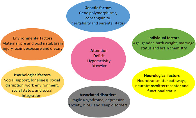
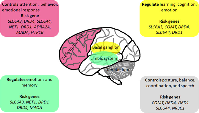
Attention-deficit hyperactivity disorder (ADHD) is a neurological and neurodevelopmental childhood-onset disorder characterized by a persistent pattern of inattentiveness, impulsiveness, restlessness, and hyperactivity. These symptoms may continue in 55-66% of cases from childhood into adulthood. Even though the precise etiology of ADHD is not fully understood, it is considered as a multifactorial and heterogeneous disorder with several contributing factors such as heritability, auxiliary to neurodevelopmental issues, severe brain injuries, neuroinflammation, consanguineous marriages, premature birth, and exposure to environmental toxins. Neuroimaging and neurodevelopmental assessments may help to explore the possible role of genetic variations on ADHD neuropsychobiology. Multiple genetic studies have observed a strong genetic association with various aspects of neuropsychobiological functions, including neural abnormalities and delayed neurodevelopment in ADHD. The advancement in neuroimaging and molecular genomics offers the opportunity to analyze the impact of genetic variations alongside its dysregulated pathways on structural and functional derived brain imaging phenotypes in various neurological and psychiatric disorders, including ADHD. Recently, neuroimaging genomic studies observed a significant association of brain imaging phenotypes with genetic susceptibility in ADHD. Integrating the neuroimaging-derived phenotypes with genomics deciphers various neurobiological pathways that can be leveraged for the development of novel clinical biomarkers, new treatment modalities as well as therapeutic interventions for ADHD patients. In this review, we discuss the neurobiology of ADHD with particular emphasis on structural and functional changes in the ADHD brain and their interactions with complex genomic variations utilizing imaging genetics methodologies. We also highlight the genetic variants supposedly allied with the development of ADHD and how these, in turn, may affect the brain circuit function and related behaviors. In addition to reviewing imaging genetic studies, we also examine the need for complementary approaches at various levels of biological complexity and emphasize the importance of combining and integrating results to explore biological pathways involved in ADHD disorder. These approaches include animal models, computational biology, bioinformatics analyses, and multimodal imaging genetics studies.


Conflict of interest statement

The authors declare no competing interests.

Figures

Fig. 1
Fig. 1. Factors affecting ADHD pathophysiology.
Different factors such as genetic, in particular, gene polymorphisms, environmental factors, psychological factors, individual factors such as age, abnormality in various neurological pathways such as dopaminergic and serotonergic, and comorbidity with multiple disorders are associated with symptoms of attention-deficit hyperactivity disorder.
Fig. 2
Fig. 2. Risk genes and associated altered brain regions in attention-deficit hyperactivity disorder.
Multiples genes are associated with altered structural and functional brain changes, mainly in the frontal lobe, basal ganglion, limbic system, and cerebellum.
Fig. 3
Fig. 3. Major pathways related to the pathogenesis of attention-deficit hyperactivity disorder (dopaminergic and serotonergic).
Dopaminergic and serotonergic neurons are primarily located respectively in the midbrain and hindbrain and control various functions. Anomalies in dopamine and/or serotonin levels contribute to the symptoms of inattention, hyperactivity, and impulsiveness in attention-deficit hyperactivity disorder (Figure inspired from the manuscript by Fontana BD et al., 2019).
Fig. 4
Fig. 4. Interaction map of hot genes associated with various parts of brain changes as observed on structural and functional MRI in attention-deficit hyperactivity disorder, generated using STRING1 webserver.
All sources are used to create the interaction model with a default medium confidence interaction score of 0.4. and k-means clustering method. The line color indicates the type of interaction evidence: blue line denotes co-occurrence, black line indicates co-expression, and the purple line indicates experimental evidence ref: https://string-db.org/.
Fig. 5
Fig. 5. Enrichment analysis of hot genes which predispose to ADHD.
Summary of the top 20 gene ontology (GO) in terms of biological processes (A), cellular components (B), and molecular functions (C). The proportion represents the number of genes enriched in each GO category. Significant enrichment genes belong to neurotransmitters (A), neuron projections, part of neurons (B), and ammonium ion (C), which play a vital role in synaptic interactions suggesting the risk factor for ADHD is polymorphism in the enriched genes.
Fig. 6
Fig. 6. Gene enrichment analysis of hot genes associated with ADHD.
Gene expression heatmaps constructed with GTEX v8 (54 tissue types). The heatmap indicates the significance of expressed gene modules related to brain regions. Blue to red reflects a significant association of the gene with brian regions as determined by a standardized z score. The gene expression heatmap showing higher relative expression levels of MAOB, SNAP25, COMT, MAOA, ADRA2C, DRD1, DRD2, HTR2C, CHRNA4, and TH in different brain sites suggest that these genes may be linked with brain areas and are considered as a risk factor for ADHD.
Fig. 7
Fig. 7. Differently expressed gene (DEG) plots of 24 ADHD hot genes constructed with GTEX v8 across 54 tissue samples.
Significantly enriched differently expressed gene sets, highlighted in red, belong to the hypothalamus and substantia nigra.
Fig. 8
Fig. 8. The figure shows structural and functional brain changes associated with gene polymorphisms in patients with ADHD.
A SLC6A3 polymorphisms are associated with lower caudate nucleus volume and prefrontal cortex in patients with ADHD. B SLC6A3 and SLC6A4 polymorphisms associated with lower functional activity in the prefrontal cortex and cerebellum in the brain of ADHD (Figure inspired from the manuscript by Tripp G, et al., 2009).

References

    1. Polanczyk G, Rohde LA. Epidemiology of attention-deficit/hyperactivity disorder across the lifespan. Curr. Opin. Psychiatry. 2007;20:386–392. doi: 10.1097/YCO.0b013e3281568d7a. - DOI - PubMed
    1. Simon V, Czobor P, Balint S, Meszaros A, Bitter I. Prevalence and correlates of adult attention-deficit hyperactivity disorder: meta-analysis. Br. J. Psychiatry.: J. Ment. Sci. 2009;1:204–211. doi: 10.1192/bjp.bp.107.048827. - DOI - PubMed
    1. Nigg JT. Attention deficits and hyperactivity-impulsivity: what have we learned, what next? Dev. Psychopathol. 2013;25:1489–503. doi: 10.1017/S0954579413000734. - DOI - PMC - PubMed
    1. Wilens TE, Spencer TJ. Understanding attention-deficit/hyperactivity disorder from childhood to adulthood. Postgrad. Med. 2010;122:97–109. doi: 10.3810/pgm.2010.09.2206. - DOI - PMC - PubMed
    1. Buitelaar JK, et al. Long-term efficacy and safety outcomes with OROS-MPH in adults with ADHD. Int. J. Neuropsychopharmacol. 2012;15:1–13. doi: 10.1017/S1461145711001131. - DOI - PMC - PubMed

Publication types