. 2021 Aug;210(4):197-209.
doi: 10.1007/s00430-021-00713-6. Epub 2021 Jun 6.

Transmission of cell-associated human cytomegalovirus isolates between various cell types using polymorphonuclear leukocytes as a vehicle

Affiliations

Transmission of cell-associated human cytomegalovirus isolates between various cell types using polymorphonuclear leukocytes as a vehicle

Berenike Braun et al. Med Microbiol Immunol. 2021 Aug.

Abstract

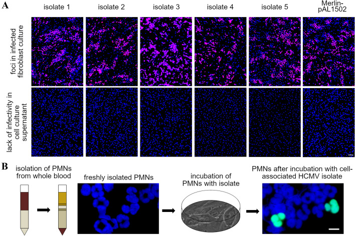
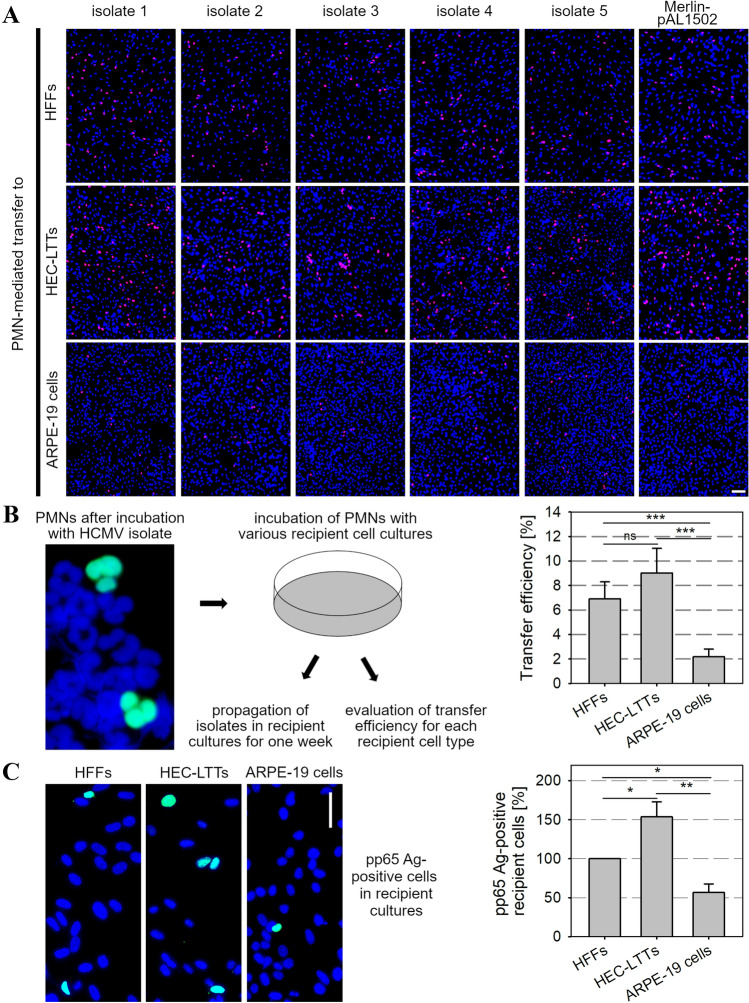
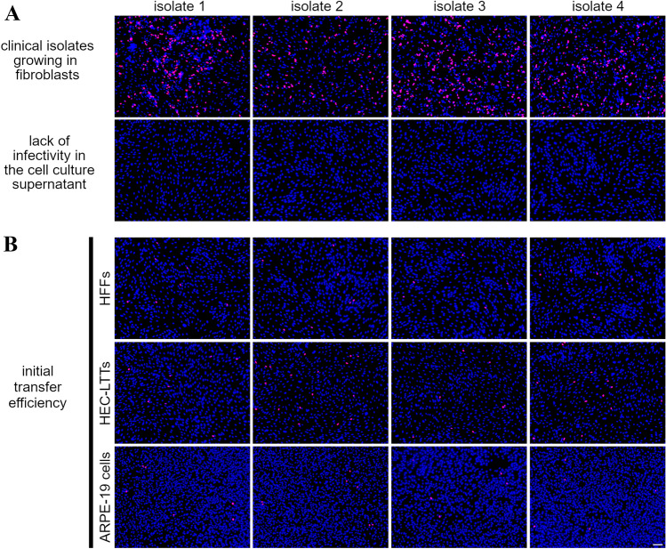
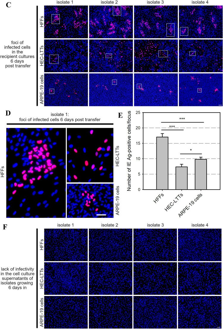
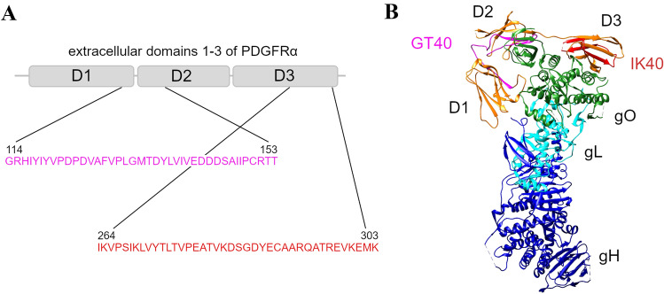
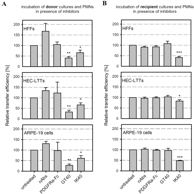
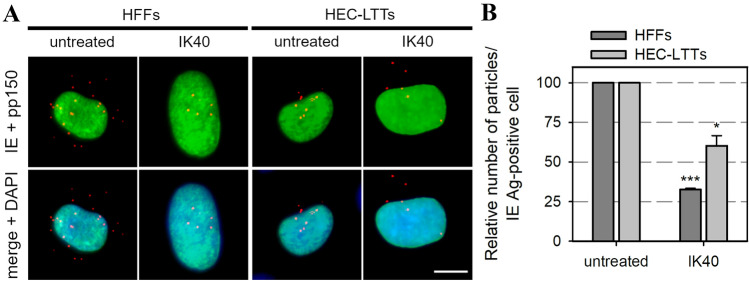
Polymorphonuclear leukocytes (PMNs) are regarded as vehicles for the hematogenous dissemination of human cytomegalovirus (HCMV). In cell culture, this concept has been validated with cell-free laboratory strains but not yet with clinical HCMV isolates that grow strictly cell-associated. We, therefore, aimed to evaluate whether PMNs can also transmit such isolates from initially infected fibroblasts to other cell types, which might further clarify the role of PMNs in HCMV dissemination and provide a model to search for potential inhibitors. PMNs, which have been isolated from HCMV-seronegative individuals, were added for 3 h to fibroblasts infected with recent cell-associated HCMV isolates, then removed and transferred to various recipient cell cultures. The transfer efficiency in the recipient cultures was evaluated by immunofluorescence staining of viral immediate early antigens. Soluble derivatives of the cellular HCMV entry receptor PDGFRα were analyzed for their potential to interfere with this transfer. All of five tested HCMV isolates could be transferred to fibroblasts, endothelial and epithelial cells with transfer rates ranging from 2 to 9%, and the transferred viruses could spread focally in these recipient cells within 1 week. The PDGFRα-derived peptides IK40 and GT40 reduced transfer by 40 and 70% when added during the uptake step. However, when added during the transfer step, only IK40 was effective, inhibiting transmission by 20% on endothelial cells and 50-60% on epithelial cells and fibroblasts. These findings further corroborate the assumption of cell-associated HCMV dissemination by PMNs and demonstrate that it is possible to inhibit this transmission mode.

Keywords: Cell-associated spread; Clinical isolates; HCMV; Hematogenous dissemination; PDGFRα-peptides; PMNs.


Conflict of interest statement

Aspects of this work are the subject of the patent filing WO2018002081A1.

Figures

Fig. 1
Fig. 1
PMNs take up cell-associated HCMV isolates from infected HFFs. A Uninfected HFFs were either co-cultured with HFFs infected with 5 different clinical isolates or infected with Merlin-pAL1502. These cultures were used as donor cultures 4–7 d p. i. (upper panel). Their cell culture supernatants were analyzed for cell-free infectivity on uninfected HFFs on the day of each transfer experiment (lower panel). Cells were fixed on the next day and stained via indirect immunofluorescence for viral IE Ag (pink nuclei). Cell nuclei were counterstained with DAPI (blue nuclei). Scale bar = 100 µm. B PMNs isolated from fresh blood samples of HCMV-seronegative donors were either directly stained for viral pp65 Ag (green nuclei) or after incubation with the HCMV donor cultures for 3 h at 37 °C. Cell nuclei were counterstained with DAPI (blue nuclei)
Fig. 2
Fig. 2
PMNs transfer cell-associated HCMV isolates with different susceptibility to various recipient cell cultures. A PMNs were recollected after incubation with the donor cultures for 3 h at 37 °C and applied onto uninfected HFFs, HEC-LTTs or ARPE-19 cells. After 3 h, PMNs were removed. On the next day, recipient cells were fixed and stained for viral IE Ag via indirect immunofluorescence (pink nuclei). Cell nuclei were counterstained with DAPI (blue nuclei). Scale bar = 100 µm. B Schematic of the transfer procedure and determination of transfer efficiencies onto the recipient cultures as the ratio of infected cells to the total cell number. Error bars represent the standard error of the mean (SEM) of 11 individual experiments, asterisks indicate significant differences (***p value < 0.001). C PMN-mediated transfer was performed with 3 different clinical isolates. On the next day, recipient cells were fixed and stained for viral pp65 Ag via indirect immunofluorescence (green nuclei). Cell nuclei were counterstained with DAPI (blue nuclei). Scale bar = 50 µm. The percentage of pp65 Ag-positive recipient cells was determined as a ratio of total cell number. Error bars represent the SEM of 3 individual experiments, asterisks indicate significant differences (*p value < 0.05; **p value < 0.01)
Fig. 3
Fig. 3
Clinical isolates grow in a focal fashion in the recipient cultures after being transferred from infected HFFs via PMNs. A Uninfected HFFs were co-cultured with HFFs infected with 4 different clinical isolates. After 2 days, these cultures were used as donor cultures for PMN-mediated transmission. The cell culture supernatants of the isolates were analyzed for cell-free infectivity on uninfected HFFs at the day of the transfer experiment. PMNs isolated from fresh blood samples of HCMV-seronegative donors were incubated with the donor cultures for 3 h at 37 °C, then recollected and incubated with uninfected HFFs, HEC-LTTs or ARPE-19 cells. After 3 h at 37 °C, PMNs were removed. B Recipient cell cultures were fixed on the next day and stained for viral IE Ag via indirect immunofluorescence (pink nuclei). Cell nuclei were counterstained with DAPI (blue nuclei). Scale bar = 100 µm. C Replica cultures were incubated for 6 days to allow growth of the isolates in the recipient cells and then fixed and stained for viral IE Ag (pink nuclei). Cell nuclei were counterstained with DAPI (blue nuclei). Scale bar = 100 µm. In each immunofluorescence image, one exemplary focus is framed. D All exemplary frames of isolate 1 are shown magnified. Scale bar = 50 µm. E The recipient cultures were evaluated for the number of IE Ag-positive cells per focus 6 days post-transfer. For each cell type, 70 foci were analyzed. Error bars represent the standard error of the mean of 4 individual experiments, asterisks indicate significant differences (*p < 0.05; ***p < 0.001). F Cell culture supernatants of isolates growing for 6 days in the recipient cultures were analyzed for cell-free infectivity on uninfected HFFs. Cell nuclei were counterstained with DAPI (blue nuclei). Scale bar = 100 µm
Fig. 3
Fig. 3
Clinical isolates grow in a focal fashion in the recipient cultures after being transferred from infected HFFs via PMNs. A Uninfected HFFs were co-cultured with HFFs infected with 4 different clinical isolates. After 2 days, these cultures were used as donor cultures for PMN-mediated transmission. The cell culture supernatants of the isolates were analyzed for cell-free infectivity on uninfected HFFs at the day of the transfer experiment. PMNs isolated from fresh blood samples of HCMV-seronegative donors were incubated with the donor cultures for 3 h at 37 °C, then recollected and incubated with uninfected HFFs, HEC-LTTs or ARPE-19 cells. After 3 h at 37 °C, PMNs were removed. B Recipient cell cultures were fixed on the next day and stained for viral IE Ag via indirect immunofluorescence (pink nuclei). Cell nuclei were counterstained with DAPI (blue nuclei). Scale bar = 100 µm. C Replica cultures were incubated for 6 days to allow growth of the isolates in the recipient cells and then fixed and stained for viral IE Ag (pink nuclei). Cell nuclei were counterstained with DAPI (blue nuclei). Scale bar = 100 µm. In each immunofluorescence image, one exemplary focus is framed. D All exemplary frames of isolate 1 are shown magnified. Scale bar = 50 µm. E The recipient cultures were evaluated for the number of IE Ag-positive cells per focus 6 days post-transfer. For each cell type, 70 foci were analyzed. Error bars represent the standard error of the mean of 4 individual experiments, asterisks indicate significant differences (*p < 0.05; ***p < 0.001). F Cell culture supernatants of isolates growing for 6 days in the recipient cultures were analyzed for cell-free infectivity on uninfected HFFs. Cell nuclei were counterstained with DAPI (blue nuclei). Scale bar = 100 µm
Fig. 4
Fig. 4
GT40 and IK40 are 40-mer peptides derived from the extracellular domain of PDGFRα. A Schematic organization of domains 1–3 of human PDGFRα including the localization and amino acid sequences of the HCMV inhibitory peptides GT40 and IK40. B Visualization of the trimeric gH/gL/gO complex of HCMV bound to PDGFRα domains 1–3 [39]. Localizations of GT40 and IK40 in PDGFRα are highlighted
Fig. 5
Fig. 5
The PDGFRα-derived peptides GT40 and IK40 inhibit PMN-mediated transmission of HCMV. PMNs isolated from fresh blood samples of HCMV-seronegative donors were added to HFFs that were infected with 5 different clinical isolates or Merlin-pAL1502. After 3 h at 37 °C, PMNs were applied onto uninfected HFFs, HEC-LTTs or ARPE-19 cells. After 3 h, PMNs were removed from the recipient cell cultures. To evaluate their effect on PMN-mediated spread, HCMV entry inhibitors were included during incubation of PMNs with donor cultures (A) or recipient cultures (B). nAbs, a soluble PDGFRα-Fc chimera or PDGFRα-derived peptides (GT40, IK40) were added at 0.5 mg/ml, 120 ng/ml or 0.45 mg/ml, respectively. Cells were fixed on the next day and stained via indirect immunofluorescence for viral IE Ag. Cell nuclei were counterstained with DAPI. Transfer efficiencies in the presence of inhibitors were calculated and are shown relative to the transfer efficiency of untreated cells. Error bars represent the standard error of the mean of 3 (A) or 11 (B) individual experiments, asterisks indicate significant differences as compared to untreated transfer (*p value < 0.05; **p value < 0.01; ***p value < 0.001)
Fig. 6
Fig. 6
The PDGFRα-derived peptide IK40 reduces the number of transferred virus particles. PMN-mediated transfer was performed with cell-associated HCMV isolates onto HFFs and HEC-LTTs in presence or absence of the inhibitory peptide IK40 (0.45 mg/ml) during incubation of PMNs and recipient cells. A The recipient cells were fixed on the next day and stained via indirect immunofluorescence for viral IE Ag (green nuclei). Capsid-associated tegument protein pp150 was stained for detection of virus particles (red dot-like signals). Cell nuclei were counterstained with DAPI (blue nuclei). Scale bar = 10 µm. B The number of virus particles per infected cell in presence of IK40 was determined and is shown relative to the number of virus particles on untreated cells (70 cells per condition). Error bars represent the standard error of the mean of 3 individual experiments, asterisks indicate significant differences as compared to untreated transfer (*p value < 0.05; ***p value < 0.001)

Similar articles

Cited by

References

    1. Pass RF, Zhang C, Evans A, Simpson T, Andrews W, Huang ML, Corey L, Hill J, Davis E, Flanigan C, Cloud G. Vaccine prevention of maternal cytomegalovirus infection. N Engl J Med. 2009;19(360):1191–1199. doi: 10.1056/NEJMoa0804749. - DOI - PMC - PubMed
    1. Britt WJ. Congenital human cytomegalovirus infection and the enigma of maternal immunity. J Virol. 2017 doi: 10.1128/JVI.02392-16. - DOI - PMC - PubMed
    1. Stagno S, Pass RF, Cloud G, Britt WJ, Henderson RE, Walton PD, Veren DA, Page F, Alford CA. Primary cytomegalovirus infection in pregnancy: incidence, transmission to fetus, and clinical outcome. JAMA. 1986;256(14):1904–1908. doi: 10.1001/jama.1986.03380140074025. - DOI - PubMed
    1. Kotton CN, Kumar D, Caliendo AM, Åsberg A, Chou S, Danziger-Isakov L, Humar A. Updated international consensus guidelines on the management of cytomegalovirus in solid-organ transplantation. Transplant J. 2013;96(4):333–360. doi: 10.1097/TP.0b013e31829df29d. - DOI - PubMed
    1. Kagan KO, Hamprecht K. Cytomegalovirus infection in pregnancy. Arch Gynecol Obstet. 2017;296(1):15–26. doi: 10.1007/s00404-017-4380-2. - DOI - PubMed

MeSH terms