. 2021 Dec;250(12):1796-1809.
doi: 10.1002/dvdy.380. Epub 2021 Jun 16.

A show of Hands: Novel and conserved expression patterns of teleost hand paralogs during craniofacial, heart, fin, peripheral nervous system and gut development

Affiliations

A show of Hands: Novel and conserved expression patterns of teleost hand paralogs during craniofacial, heart, fin, peripheral nervous system and gut development

Samantha Reynolds et al. Dev Dyn. 2021 Dec.

Abstract

Background: Hand genes are required for the development of the vertebrate jaw, heart, peripheral nervous system, limb, gut, placenta, and decidua. Two Hand paralogues, Hand1 and Hand2, are present in most vertebrates, where they mediate different functions yet overlap in expression. In ray-finned fishes, Hand gene expression and function is only known for the zebrafish, which represents the rare condition of having a single Hand gene, hand2. Here we describe the developmental expression of hand1 and hand2 in the cichlid Copadichromis azureus.

Results: hand1 and hand2 are expressed in the cichlid heart, paired fins, pharyngeal arches, peripheral nervous system, gut, and lateral plate mesoderm with different degrees of overlap.

Conclusions: Hand gene expression in the gut, peripheral nervous system, and pharyngeal arches may have already been fixed in the lobe- and ray-finned fish common ancestor. In other embryonic regions, such as paired appendages, hand2 expression was fixed, while hand1 expression diverged in lobe- and ray-finned fish lineages. In the lateral plate mesoderm and arch associated catecholaminergic cells, hand1 and hand2 swapped expression between divergent lineages. Distinct expression of cichlid hand1 and hand2 in the epicardium and myocardium of the developing heart may represent the ancestral pattern for bony fishes.

Keywords: cichlid; epicardium; fin; gut; hand1; hand2; heart; myocardium; oral teeth; pharyngeal arches; sympathetic.


Conflict of interest statement

CONFLICT OF INTEREST

The authors declare that they have no competing interests.

Figures
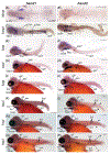
FIGURE 1
FIGURE 1
hand1 and hand2 expression in 2 to 8dpf C. azureus. A-B: hand1, A, and hand2, B, expression at 36 hpf in de-yolked embryos (dorsal view). hand1 expression is detected in anterior and posterior lateral plate mesoderm, while hand2 is not. C-D, hand1, C, and hand2, D, expression in 54 hpf de-yolked embryos (ventral view). hand1 is detected in the heart, lateral plate mesoderm and gut, C, and hand2 expression is detected in the heart and pharyngeal arches, D. E and F, hand1, E, and hand2, F, expression in 3 dpf de-yolked embryos (lateral view). hand1 and hand2 expression is detected in the heart, pharyngeal arches, and pectoral fins. hand1 alone is expressed in the gut, E. G and H, hand1, G, and hand2, H, expression at 4 dpf. hand1 and hand2 are still detected in the heart, pharyngeal arches, and pectoral fins. hand2 is first detected in the gut, H. I and J, hand1, I, and hand2, J, expression at 5 dpf. hand1 and hand2 are still detected in the heart, pharyngeal arches, pectoral fins, and gut. Their domains of expression now include vagal ganglia (gX). hand1 pharyngeal arch expression has become restricted to the mandibular and hyoid arches, I. K and L, hand1, K, and hand2, L, expression at 6 dpf. hand1 and hand2 are still detected in the heart, pharyngeal arches, pectoral fins, gut, and vagal ganglia. hand1 and hand2 are first detected in trunk sympathetic ganglia. hand2 is first detected in the presumptive dorsal and anal fins (arrowheads), L. M and N, hand1, M, and hand2, N, expression at 7 dpf. hand1 and hand2 are still detected in the heart, pharyngeal arches, pectoral fins, gut, vagal, and trunk sympathetic ganglia, and in the dorsal and anal fins. O and P, hand1, O, and hand2, P, expression at 8 dpf. hand1 and hand2 are still detected in the heart, pharyngeal arches, pectoral fins, gut, vagal and trunk sympathetic ganglia, and in the dorsal and anal fins. Their domains of expression include pelvic fins starting at this stage. Anterior to the left. * indicates first detection stage in labeled structure. aLPM, anterior lateral plate mesoderm; pLPM, posterior lateral plate mesoderm; PA, pharyngeal arches; pec, pectoral fin; SG, sympathetic ganglia; ma, mandibular arch; hy, hyoid arch; gX, vagus ganglion; df, dorsal fin; af, anal fin; pel, pelvic fin. Scale: 100 μm
FIGURE 2
FIGURE 2
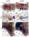
hand1 and hand2 expression in the developing cichlid heart. A and B, hand1, A, and hand2, B, expression in the 54 hpf heart (ventral view). hand1 expression is detected in the outer heart layer, which we interpret as epicardium (ep), while hand2 expression is detected in the inner heart layer, which we interpret as presumptive myocardium (my). hand1 is also expressed in arch associated catecholaminergic cells (arrowhead). C and D, hand1 C, and hand2, D, expression in the 78 hpf dissected heart (ventral view). C, hand1 expression is detected in the epicardium of ventricle, outflow tract, and proximal atrium. D, hand2 expression is robust in the myocardium of the ventricle and outflow tract, and weak in the atrium. E and F, hand1 (E) and hand2 (F) expression in the 7 dpf heart (lateral view). E, hand1 is expressed in the outer layers of the ventricle and outflow tract and not detected in the atrium. F, hand2 is expressed in the inner layers of the atrium, ventricle, and outflow tract. Both genes are also expressed in the cardiac sac. at, atrium; cs, cardiac sac; oft, outflow tract; ve, ventricle. Scale: 25 μm
FIGURE 3
FIGURE 3
hand1 and hand2 expression in the developing cichlid paired and median fins. A and B, hand1, A, and hand2, B, expression in the 3 dpf pectoral fin. hand1, A, is expressed in the entire fin mesenchyme, while hand2, B, is expressed in the posterior mesenchyme. C and D, hand1, C, and hand2, D, expression in the 7 dpf pectoral fin (anterior to the left). hand1, C, fin base expression is continuous along the A-P axis, while hand2, D, expression is restricted to the posterior half of the fin base. E and F, hand1, E, and hand2, F, expression in the 9 dpf pelvic fin. hand1, E, is expressed in the entire fin mesenchyme, while hand2, F, is expressed in the posterior mesenchyme. G and H, hand2, H, but not hand1, G, is expressed in the posterior mesenchyme of the 9 dpf dorsal fin. I and J: hand2, I, but not hand1, J, is expressed in the posterior mesenchyme of the 9 dpf anal fin. Scale: 25 μm. pec, pectoral fin; pel, pelvic fin; df, dorsal fin; af, anal fin
FIGURE 4
FIGURE 4
hand1 and hand2 expression in the developing cichlid gut. A, hand1 expression throughout the 7 dpf intestinal mesenchyme. B, hand2 salt and pepper expression in the 7 dpf intestinal mesenchyme. C, sagittal section showing gut histology at 7dpf. Scale: 50 μm
FIGURE 5
FIGURE 5
hand1 and hand2 expression in the developing cichlid pharyngeal arches. A and B, hand1, A, and hand2, B, overlapping expression in the 3 dpf pharyngeal arches. hand1, A, expression is more restricted than hand1 to the ventral-most domain. C to F: hand1 (C,E) and hand2 (D,F) expression in the 7 dpf pharyngeal arches. hand1 (C, lateral; E, ventral) expression is restricted to pharyngeal arches 1 to 2, while hand2 (D, lateral; F, ventral) is expressed in all arches. G and H: hand1, G, and hand2, H, expression is not detected in the 9 dpf upper jaw dental mesenchyme. Dorsal to the top. I and J, hand1, I, and hand2, J, expression in the 9 dpf lower jaw. hand1, I, expression is strongest at the midline (left in image), and in the dental mesenchyme. hand2 expression is detected in the mesenchyme posterior Meckel’s cartilage. Neither gene is detected in Meckel’s cartilage. Anterior to the top. K and L, hand1, K, and hand2, L, expression in the 9 dpf hyoid arch. hand1 and hand2 are both expressed in midline mesenchyme in the region connecting to the cardiac outflow tract. hand2 is also expressed in mesenchyme around the hypo- and cerato-hyal cartilages, L, Anterior to the top. M to P, hand1 (M,O) and hand2 (N,P) expression is not detected in the 9 dpf upper (M,N) and lower (O,P) pharyngeal jaw dental mesenchyme. Anterior to the top. ch, ceratohyal; gX, vagal ganglion; hh, hypohyal; Mc, Meckel’s cartilage; PA, pharyngeal arch; pec, pectoral fin. Scale: 50 μm
FIGURE 6
FIGURE 6
hand1 and hand2 expression in the developing cichlid PNS. A to C, hand1, A, and hand2, B, expression in sympathetic ganglia at 7 dpf. C, sagittal section showing sympathetic ganglia at 7 dpf (arrows). D to I, hand1 and hand2 are expressed in the vagal ganglion. D and G, hand1 expression in the vagal ganglion in lateral view. E and H, hand2 expression in the vagal ganglion in lateral view. F and I, sagittal section showing vagal ganglion histology. J to L, hand1, J, and hand2, K, expression in the peripheral nervous system extends into the head region and labels the left and right sympathetic ganglion chains, L. Scale: 50 μm. eb1-4, epibranchial 1-4; gT1, first trunk sympathetic ganglion; gX, vagal ganglion; k, kidney; my, myotome; not, notochord; pec, pectoral fin; SG, sympathetic ganglion; SN, sympathetic nerve; th, thymus

References

    1. Massari ME, Murre C. Helix-loop-helix proteins: regulators of transcription in eucaryotic organisms. Mol Cell Biol. 2000;20(2):429–440. 10.1128/mcb.20.2.429-440.2000. - DOI - PMC - PubMed
    1. Moore AW, Barbel S, Jan LY, Jan YN. A genomewide survey of basic helix-loop-helix factors in drosophila. Proc Natl Acad Sci U S A. 2000;97(19):10436–10441. 10.1073/pnas.170301897. - DOI - PMC - PubMed
    1. Kolsch V, Paululat A. The highly conserved cardiogenic bHLH factor hand is specifically expressed in circular visceral muscle progenitor cells and in all cell types of the dorsal vessel during drosophila embryogenesis. Dev Genes Evol. 2002;212(10):473–485. 10.1007/s00427-002-0268-6. - DOI - PubMed
    1. Han Z, Olson EN. Hand is a direct target of Tinman and GATA factors during drosophila cardiogenesis and hematopoiesis. Development. 2005;132(15):3525–3536. 10.1242/dev.01899. - DOI - PubMed
    1. Holland LZ, Ocampo DD. A new look at an old question: when did the second whole genome duplication occur in vertebrate evolution? Genome Biol. 2018;19209:209. 10.1186/s13059-018-1592-0. - DOI - PMC - PubMed

Publication types

MeSH terms

Substances