. 2021 Aug;21(8):2785-2794.
doi: 10.1111/ajt.16708. Epub 2021 Jun 25.

T cell-mediated response to SARS-CoV-2 in liver transplant recipients with prior COVID-19

Affiliations

T cell-mediated response to SARS-CoV-2 in liver transplant recipients with prior COVID-19

Mario Fernández-Ruiz et al. Am J Transplant. 2021 Aug.

Abstract

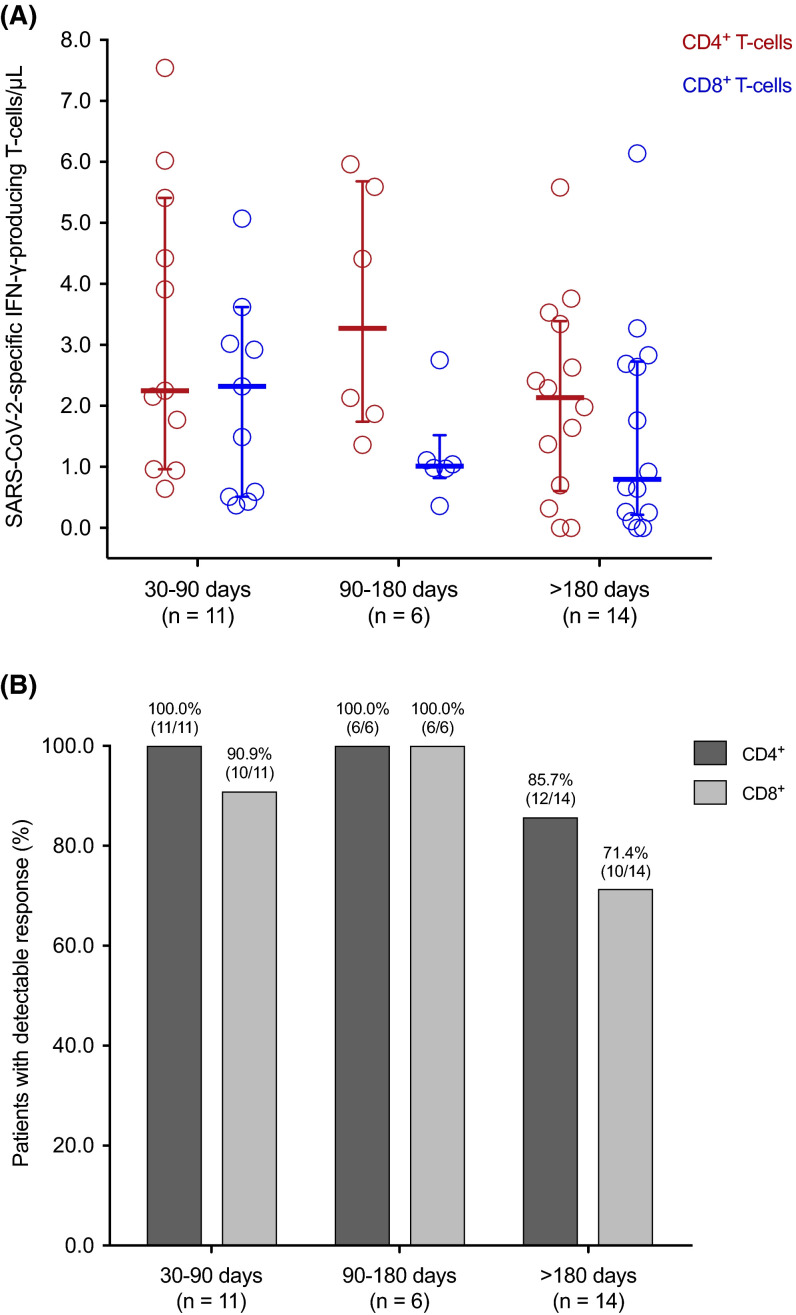
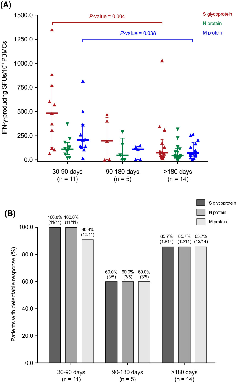
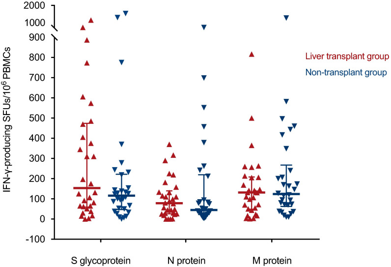
Whether immunosuppression impairs severe acute respiratory syndrome coronavirus 2-specific T cell-mediated immunity (SARS-CoV-2-CMI) after liver transplantation (LT) remains unknown. We included 31 LT recipients in whom SARS-CoV-2-CMI was assessed by intracellular cytokine staining (ICS) and interferon (IFN)-γ FluoroSpot assay after a median of 103 days from COVID-19 diagnosis. Serum SARS-CoV-2 IgG antibodies were measured by ELISA. A control group of nontransplant immunocompetent patients were matched (1:1 ratio) by age and time from diagnosis. Post-transplant SARS-CoV-2-CMI was detected by ICS in 90.3% (28/31) of recipients, with higher proportions for IFN-γ-producing CD4+ than CD8+ responses (93.5% versus 83.9%). Positive spike-specific and nucleoprotein-specific responses were found by FluoroSpot in 86.7% (26/30) of recipients each, whereas membrane protein-specific response was present in 83.3% (25/30). An inverse correlation was observed between the number of spike-specific IFN-γ-producing SFUs and time from diagnosis (Spearman's rho: -0.418; p value = .024). Two recipients (6.5%) failed to mount either T cell-mediated or IgG responses. There were no significant differences between LT recipients and nontransplant patients in the magnitude of responses by FluoroSpot to any of the antigens. Most LT recipients mount detectable-but declining over time-SARS-CoV-2-CMI after a median of 3 months from COVID-19, with no meaningful differences with immunocompetent patients.

Keywords: clinical research/practice; complication: infectious; immunosuppression/immune modulation; infection and infectious agents; infection and infectious agents - viral; infectious disease; liver transplantation/hepatology; monitoring: immune.


Figures

FIGURE 1
FIGURE 1
SARS-CoV-2-specific IFN-γ-producing T cell CD4+ (red) and CD8+ (blue) T cell counts (A) and proportion of patients with detectable (≥0.1%) responses (B) by the ICS method according to the time interval from the diagnosis of COVID-19 to the assessment. Horizontal bars and whiskers represent median values and interquartile ranges, respectively. IFN-γ, interferon-γ; SARS-CoV-2, severe acute respiratory syndrome coronavirus 2
FIGURE 2
FIGURE 2
SARS-CoV-2-specific IFN-γ-producing T cell responses reactive to the S glycoprotein (red), the N protein (green), and the M protein (blue) (A) and proportion of patients with positive (>25 S-reactive, >14 N-reactive and >21 M-reactive SFUs/106 PBMCs) responses (B) by the IFN-γ FluoroSpot assay according to the interval from the diagnosis of COVID-19 to the assessment. Horizontal bars and whiskers represent median values and interquartile ranges, respectively. Comparisons were performed with the Mann-Whitney U test. IFN-γ, interferon-γ; PBMC, peripheral blood mononuclear cell; SARS-CoV-2, severe acute respiratory syndrome coronavirus 2; SFU, spot forming unit
FIGURE 3
FIGURE 3
Correlation (Spearman’s rho) between the semiquantitative results of SARS-CoV-2 IgG ELISA and the number of M protein-specific IFN-γ-producing SFUs per 106 PBMCs by the IFN-γ FluoroSpot assay. IFN-γ, interferon-γ; PBMC, peripheral blood mononuclear cell; SARS-CoV-2, severe acute respiratory syndrome coronavirus 2; SFU, spot forming unit
FIGURE 4
FIGURE 4
SARS-CoV-2-specific IFN-γ-producing T cell responses measured by the IFN-γ FluoroSpot assay in 30 LT recipients (red) and 30 nontransplant patients (blue) matched (1:1 ratio) by age and time interval from the diagnosis of COVID-19. Horizontal bars and whiskers represent median values and interquartile ranges, respectively. IFN-γ, interferon-γ; PBMC, peripheral blood mononuclear cell; SARS-CoV-2, severe acute respiratory syndrome coronavirus 2; SFU, spot forming unit

References

    1. Coll E, Fernández-Ruiz M, Sánchez-Álvarez JE, et al. COVID-19 in transplant recipients: the Spanish experience. Am J Transplant. 2021;21(5):1825–1837. doi: 10.1111/ajt.16369. - DOI - PMC - PubMed
    1. Colmenero J, Rodríguez-Perálvarez M, Salcedo M, et al. Epidemiological pattern, incidence, and outcomes of COVID-19 in liver transplant patients. J Hepatol. 2021;74:148–155. - PMC - PubMed
    1. Kates OS, Haydel BM, Florman SS, et al. COVID-19 in solid organ transplant: a multi-center cohort study [published online ahead of print]. Clin Infect Dis. 2020. 10.1093/cid/ciaa1097 - DOI - PubMed
    1. Candon S, Guerrot D, Drouot L, et al. T cell and antibody responses to SARS-CoV-2: experience from a French transplantation and hemodialysis center during the COVID-19 pandemic. Am J Transplant. 2021;21:854–863. doi: 10.1111/ajt.16348. - DOI - PMC - PubMed
    1. Fernández-Ruiz M, Olea B, Giménez E, et al. SARS-CoV-2-specific cell-mediated immunity in kidney transplant recipients recovered from COVID-19. Transplantation. 2021;105(6):1372–1380. doi: 10.1097/TP.0000000000003672. - DOI - PubMed

Publication types

Substances