

Case of Rapidly Expanding Conjunctival Malignant Melanoma Initially from Primary Acquired Melanosis Diagnosed 14 Years Earlier
- PMID: 34093043
- PMCID: PMC8168958
- DOI: 10.2147/IMCRJ.S310702
Case of Rapidly Expanding Conjunctival Malignant Melanoma Initially from Primary Acquired Melanosis Diagnosed 14 Years Earlier
Abstract
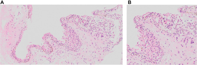
Primary acquired melanosis (PAM) of the conjunctiva is a potentially serious melanocytic lesion that can lead to the development of a melanoma. A 60-year-old woman noticed pigmentation of the conjunctiva of her left eye for more than 10 years. She underwent excisional biopsy combined with cryotherapy and was diagnosed with PAM without atypia by intraoperative consultation. She was followed for 7 years, and no changes were observed. Fourteen years after the initial biopsy, she noted a growing conjunctival tumor, and a melanoma was suspected. She underwent orbital exenteration and skin grafting procedures. Histopathological examination of the specimen led to a diagnosis of conjunctival malignant melanoma. Re-examination of the initial biopsy specimen revealed that there was a proliferation of melanocytes that partially expanded over the basal layer of the conjunctiva which had been diagnosed as PAM with moderate atypia. We conclude that this case of conjunctival PAM had progressed to a conjunctival malignant melanoma after 14 years. Pathological evaluation of intraepithelial lesions has its limitations; thus, cases of PAM, even in the absence of obvious atypia, require careful follow-up.
Keywords: conjunctival malignant melanoma; conjunctival melanoma; conjunctival tumor; malignant melanoma; primary acquired melanosis.
© 2021 Jimura et al.
Conflict of interest statement
Dr Chie Sotozono reports grants from Japan Agency for Medical Research and Development (AMED), Ministry of Health, Labour and Welfare, Japan, Japanese Ministry of Education, Culture, Sports, Science and Technology, and a Research Grant, outside the submitted work. The authors declare that they have no other competing interests.
Figures


Similar articles
-
Primary acquired melanosis of the conjunctiva: risks for progression to melanoma in 311 eyes. The 2006 Lorenz E. Zimmerman lecture.Ophthalmology. 2008 Mar;115(3):511-519.e2. doi: 10.1016/j.ophtha.2007.07.003. Epub 2007 Sep 20. Ophthalmology. 2008. PMID: 17884168
-
Primary acquired melanosis of the conjunctiva.Optometry. 2006 May;77(5):223-8. doi: 10.1016/j.optm.2006.02.007. Optometry. 2006. PMID: 16651212
-
Topical mitomycin chemotherapy for conjunctival malignant melanoma and primary acquired melanosis with atypia: clinical experience with histopathologic observations.Arch Ophthalmol. 2000 Jul;118(7):885-91. Arch Ophthalmol. 2000. PMID: 10900099 Clinical Trial.
-
Conjunctival melanoma arising from diffuse primary acquired melanosis in a young black woman.Cornea. 2005 Apr;24(3):352-5. doi: 10.1097/01.ico.0000141229.18472.a2. Cornea. 2005. PMID: 15778614 Review.
-
Management of pigmented conjunctival lesions.Ocul Surf. 2012 Oct;10(4):251-63. doi: 10.1016/j.jtos.2012.08.002. Epub 2012 Aug 11. Ocul Surf. 2012. PMID: 23084146 Review.
References
-
- Folberg R, McLean IW, Zimmerman LE. Primary acquired melanosis of the conjunctiva. Hum Pathol. 1985;16:129–135. - PubMed
-
- Shields JA, Shields CL, Mashayekhi A, et al. Primary acquired melanosis of the conjunctiva: risks for progression to melanoma in 311 eyes. The 2006 Lorenz E. Zimmerman lecture. Ophthalmology. 2008;115:511.e2–519.e2. - PubMed
Publication types
LinkOut - more resources
Full Text Sources
Miscellaneous
