. 2021 May 12:2021:5551578.
doi: 10.1155/2021/5551578. eCollection 2021.

The Severity of CVB3-Induced Myocarditis Can Be Improved by Blocking the Orchestration of NLRP3 and Th17 in Balb/c Mice

Affiliations

The Severity of CVB3-Induced Myocarditis Can Be Improved by Blocking the Orchestration of NLRP3 and Th17 in Balb/c Mice

Jifei Chen et al. Mediators Inflamm. .

Abstract

Background: The functional characteristics of NLRP3 in the pathogenesis of coxsackievirus B3- (CVB3-) induced viral myocarditis (VMC) have not been fully elucidated, and the targeted therapeutic effect of NLRP3 or its related pathway in VMC has not been reported.

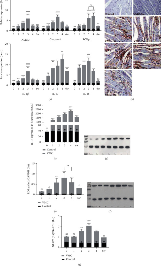
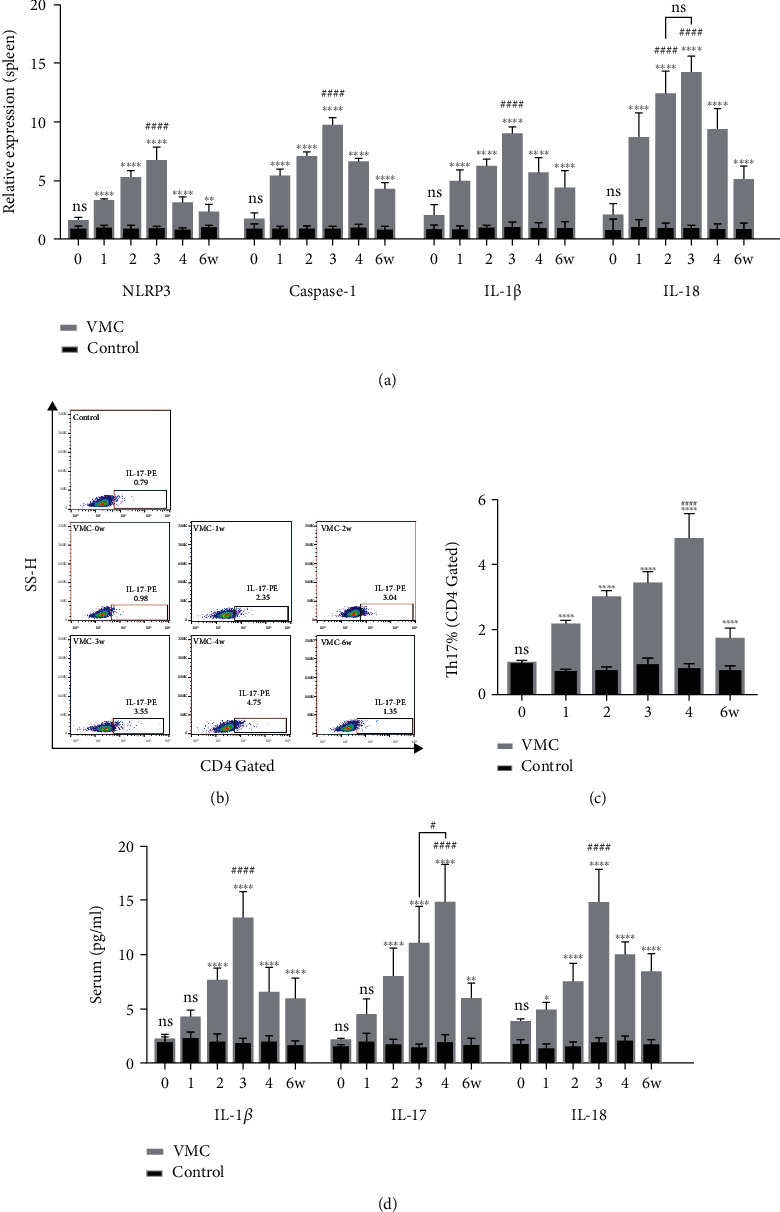
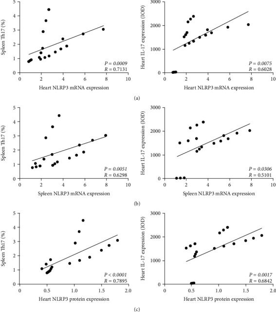
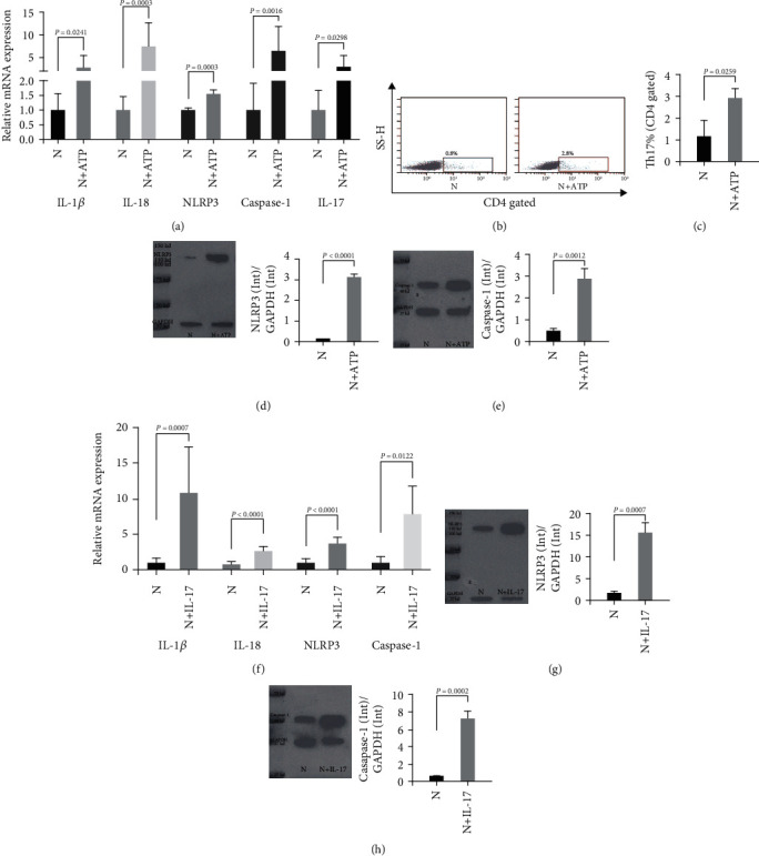
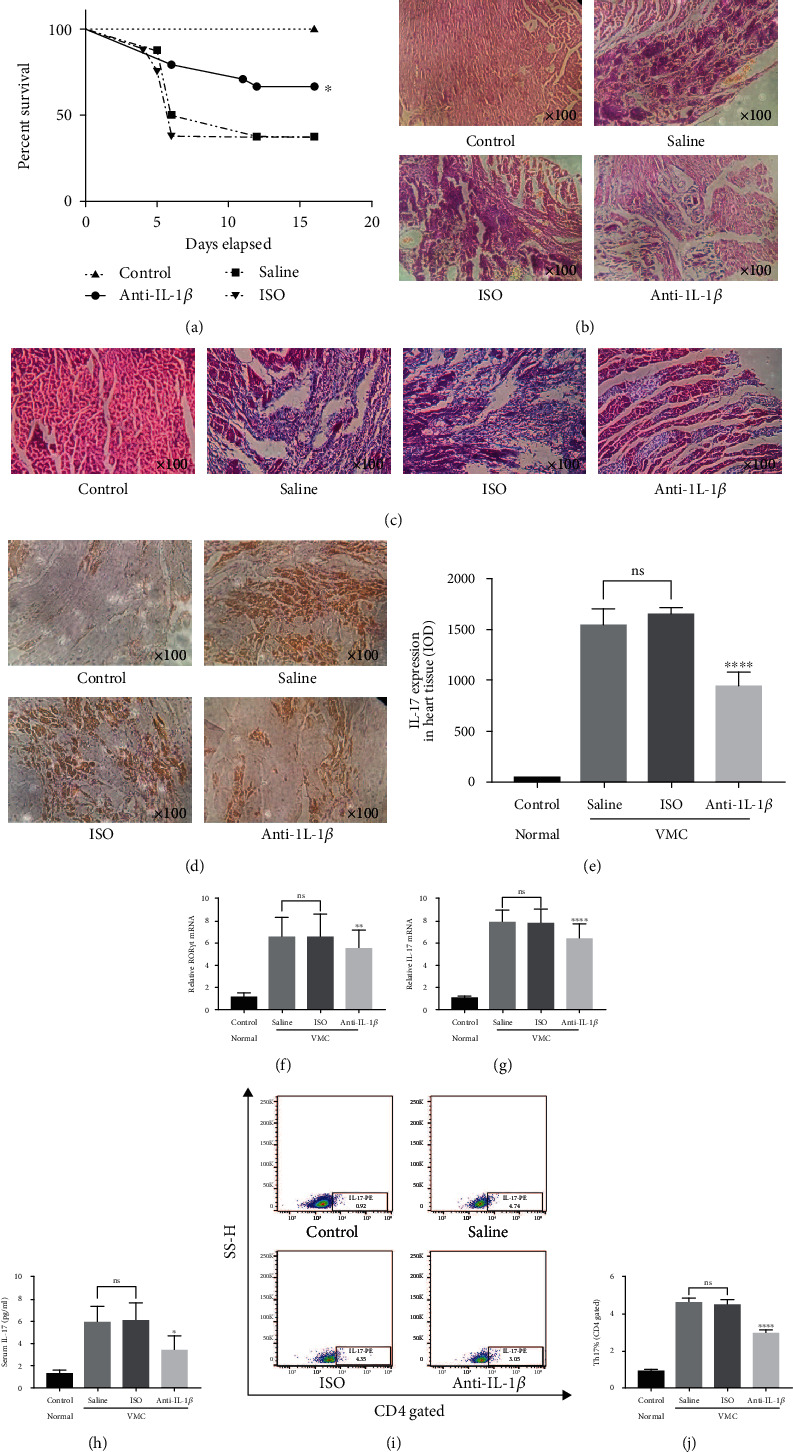
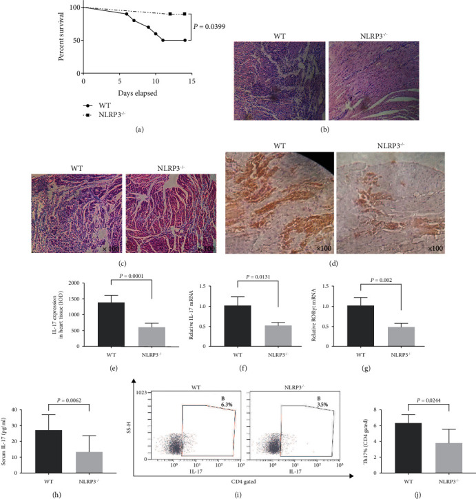
Method: In this work, the change patterns of NLRP3- and Th17-related factors were detected during the pathological process of CVB3-induced VMC in Balb/c mice. The correlation between NLRP3 and Th17 cells during the VMC process was analyzed by Spearman test. The coculture system of spleen CD4+ T and bone marrow CD11c+ DC cells was set to explore the orchestration of NLRP3 and Th17 in the pathological development of VMC in vitro. Anti-IL-1β antibody or NLRP3-/- Balb/c were used to block the NLRP3 pathway indirectly and directly to analyze the NLRP3-targeting therapeutic value.

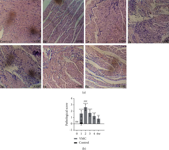
Results: The change patterns of NLRP3- and Th17-related molecules in the whole pathological process of mouse CVB3-induced VMC were described. Through Spearman correlation analysis, it was confirmed that there was a close correlation between NLRP3 and Th17 cells in the whole pathological process of VMC. And the interaction mode between NLRP3 and Th17 was preliminarily explored in the cell experiment in vitro. Under the intervention of an anti-IL-1β antibody or NLRP3 knockout, the survival rate of the intervention group was significantly improved, the degree of myocardial inflammation and fibrosis was significantly alleviated, and the content of myocardial IL-17 and spleen Th17 was also significantly decreased.

Conclusion: Our findings demonstrated a key role of the NLRP3 inflammasome and its close relationship with Th17 in the pathological progression of CVB3-induced VMC and suggested a possible positive feedback-like mutual regulation mechanism between the NLRP3 inflammasome and Th17 in vitro and in the early stage of CVB3 infection. Taking NLRP3 as a new starting point, it provides a new target and idea for the prevention and treatment of CVB3-induced VMC.


Conflict of interest statement

The authors declare that they have no competing interests.

Figures

Figure 1
Figure 1
Myocardial pathological changes in VMC process. (a) HE images of inflammation during the six-week VMC modeling process (original magnification ×100). (b) Pathological score at different time points. Statistical description: ns, ∗∗, ∗∗∗, and ∗∗∗∗ stand for no statistically significant difference, P < 0.01, P < 0.001, and P < 0.0001, respectively, for the VMC group versus the control group at different time points. ### means P < 0.001 for the week 2 VMC group versus week 0, 1, 3, 4, and 6 VMC groups.
Figure 2
Figure 2
The change pattern of NLRP3-related molecules in myocardial tissue in VMC process. (a) Relative mRNA expression of NLRP3, Caspase-1, RORγt, IL-1β, IL-17, and IL-18 in heart tissues in CVB3-induced VMC. (b) IL-17 expression in heart tissue detected by IHC (original magnification ×400). (c) IL-17 expression in heart tissue acquired by IHC assay, and IOD value was calculated by Image-Pro Plus. (d, e) The protein expression of RORγt in heart tissue was detected by WB and calculated by Bio-Rad Image Lab 6.0. (f, g) The protein expression of NLRP3 in heart tissue was detected by WB and calculated by Bio-Rad Image Lab 6.0. Statistical description: ns, ∗, ∗∗, ∗∗∗, and ∗∗∗∗ stand for no statistically significant difference, P < 0.05, P < 0.01, P < 0.001, and P < 0.0001, respectively, for VMC group versus control group at different time points. ## and ### mean P < 0.001 for the marked VMC group versus other VMC groups.
Figure 3
Figure 3
The change pattern of NLRP3-related molecules in the spleen and serum in VMC process. (a) Relative mRNA expression of NLRP3, Caspase-1, IL-1β, and IL-18 in spleen tissues during VMC process. (b) The proportion of Th17 cells in spleen samples during VMC process. (c) Statistical histogram of Th17 cell proportion calculation. (d) Protein level of IL-1β, IL-17, and IL-18 in serum during VMC process. Statistical description: ns, ∗, ∗∗, and ∗∗∗∗ stand for no statistically significant difference, P < 0.05, P < 0.01, and P < 0.0001, respectively, for VMC group versus control group at different time points. #, ###, and #### mean P < 0.05, P < 0.001, and P < 0.0001 for the marked VMC group versus other VMC groups.
Figure 4
Figure 4
Correlation analysis of NLRP3 inflammasome and Th17 cells. (a) Correlation analysis between NLRP3 (mRNA level in the heart) and Th17 cells (proportion in the spleen, left) or IL-17 expression (IOD score detected by IHC in heart, right). (b) Correlation analysis between NLRP3 (mRNA level in the spleen) and Th17 cells (proportion in the spleen, left) or IL-17 expression (IOD score detected by IHC in the heart, right). (c) Correlation analysis between NLRP3 (protein level in the heart) and Th17 cells (proportion in the spleen, left) or IL-17 expression (IOD score detected by IHC in the heart, right). P: P value; R: Spearman R.
Figure 5
Figure 5
Orchestration of NLRP3 inflammasome and Th17 cells in CVB3-induced myocarditis. (a) The relative mRNA expression of IL-1β, IL-18, NLRP3, Caspase-1, and IL-17 in N and N+ATP groups in the coculture system. (b) The proportion of Th17 cells in N and N+ATP groups in the coculture system. (c) Statistic calculation column of Th17 proportion in N and N+ATP groups in the coculture system. (d) The protein level of NLRP3 in N and N+ATP groups in the coculture system. (e) The protein level of Caspase-1 in N and N+ATP groups in the coculture system. (f) The relative mRNA levels of IL-1β, IL-18, NLRP3, and Caspase-1 in the CD11c+ DC cells stimulated with or without IL-17. (g) The protein level of NLRP3 in CD11c+ DC cells stimulated with or without IL-17. (h) The protein level of Caspase-1 in CD11c+ DC cells stimulated with or without IL-17. (a–e) were acquired from the CD11c+ DC and CD4+ T cell coculture system stimulated by LPS with (N+ATP) or without ATP (N). (f–h) were acquired from CD11c+ DC cells stimulated with IL-17 (N+IL-17) or without IL-17 (N). Statistical description: the P value in the figure has been marked between the corresponding groups.
Figure 6
Figure 6
Blockade of IL-1β can significantly alleviate the severity of VMC in Balb/c. (a) Statistical analysis of survival rate from day zero to day sixteen in control, anti-IL-1β, saline, and ISO groups. (b) HE images of myocardial in control, anti-IL-1β, saline, and ISO groups at sixteenth day of CVB3-induced VMC process (original magnification ×100). (c) Masson-trichrome staining of myocardial in control, saline, ISO, and anti-IL-1β groups at sixteenth day of CVB3-induced VMC process (original magnification ×100). (d) IHC images of IL-17 in myocardial in control, saline, ISO, and anti-IL-1β groups at sixteenth day of CVB3-induced VMC process (original magnification ×100). (e) IL-17 expression in heart tissue acquired by IHC assay and IOD value was calculated by Image-Pro Plus. (f, g) The relative mRNA expression of RORγt and IL-17 in myocardium in control, anti-IL-1β, saline, and ISO groups at sixteenth day of CVB3-induced VMC process. (h) The protein level of IL-17 in plasma in control, anti-IL-1β, saline, and ISO groups at sixteenth day of CVB3-induced VMC process. (i) The proportion of Th17 cells in spleen samples in control, anti-IL-1β, saline, and ISO groups at sixteenth day of CVB3-induced VMC process. (j) Statistical histogram of Th17 proportion in (i). Statistical description: ns, ∗, ∗∗, and ∗∗∗∗ stand for no statistically significant difference, P < 0.05, P < 0.01, and P < 0.0001, respectively, for anti-IL-1β group versus saline or ISO group.
Figure 7
Figure 7
Knockout of NLRP3 significantly alleviates the severity of CVB3-induced VMC in Balb/c. (a) Statistical analysis of survival rate from day zero to day fourteen in WT and NLRP3−/− group. (b) HE images of myocardial in WT and NLRP3−/− groups at fourteenth day of CVB3-induced VMC process (original magnification ×100 and ×200). (c) Masson-trichrome staining of myocardial in WT and NLRP3−/− groups at fourteenth day of CVB3-induced VMC process (original magnification ×100). (d) IHC images of IL-17 in myocardial in WT and NLRP3−/− groups at fourteenth day of CVB3-induced VMC process (original magnification ×100). (e) IL-17 expression in heart tissue acquired by IHC assay and IOD value was calculated by Image-Pro Plus. (f, g) The relative mRNA expression of IL-17 and RORγt in myocardium in WT and NLRP3−/− groups at fourteenth day of CVB3-induced VMC process. (h) The protein level of IL-17 in plasma in WT and NLRP3−/− groups at fourteenth day of CVB3-induced VMC process. (i) The proportion of Th17 cells in spleen samples in WT and NLRP3−/− groups at fourteenth day of CVB3-induced VMC process. (j) Statistical histogram of Th17 proportion in (i). Statistical description: the P value in the figure has been marked between the corresponding groups.

Similar articles

Cited by

References

    1. Vos T., Barber R. M., Bell B., et al. Global, regional, and national incidence, prevalence, and years lived with disability for 301 acute and chronic diseases and injuries in 188 countries, 1990-2013: a systematic analysis for the Global Burden of Disease Study 2013. The Lancet. 2015;386(9995):743–800. doi: 10.1016/s0140-6736(15)60692-4. - DOI - PMC - PubMed
    1. Esfandiarei M., McManus B. M. Molecular biology and pathogenesis of viral myocarditis. Annual Review of Pathology: Mechanisms of Disease. 2008;3(1):127–155. doi: 10.1146/annurev.pathmechdis.3.121806.151534. - DOI - PubMed
    1. Pollack A., Kontorovich A. R., Fuster V., Dec G. W. Viral myocarditis--diagnosis, treatment options, and current controversies. Nature Reviews. Cardiology. 2015;12(11):670–680. doi: 10.1038/nrcardio.2015.108. - DOI - PubMed
    1. Swanson K. V., Deng M., Ting J. P. The NLRP3 inflammasome: molecular activation and regulation to therapeutics. Nature Reviews. Immunology. 2019;19(8):477–489. doi: 10.1038/s41577-019-0165-0. - DOI - PMC - PubMed
    1. Wang Y., Gao B., Xiong S. Involvement of NLRP3 inflammasome in CVB3-induced viral myocarditis. American Journal of Physiology-Heart and Circulatory Physiology. 2014;307(10):H1438–H1447. doi: 10.1152/ajpheart.00441.2014. - DOI - PubMed

MeSH terms