. 2021 May 20:15:642819.
doi: 10.3389/fnhum.2021.642819. eCollection 2021.

Clinical Validation of the Champagne Algorithm for Epilepsy Spike Localization

Affiliations

Clinical Validation of the Champagne Algorithm for Epilepsy Spike Localization

Chang Cai et al. Front Hum Neurosci. .

Abstract

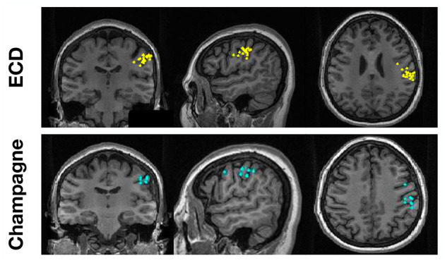
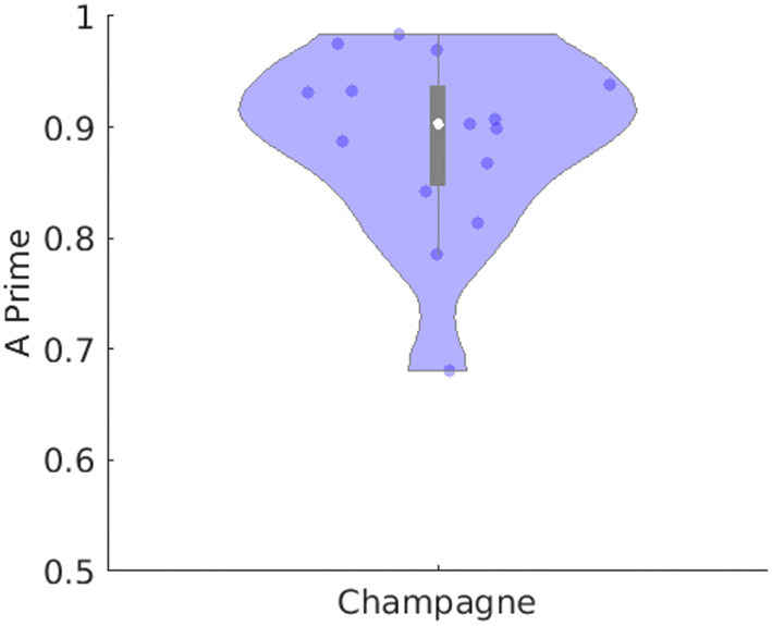
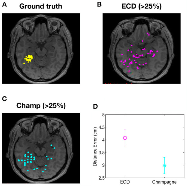
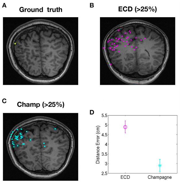
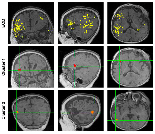
Magnetoencephalography (MEG) is increasingly used for presurgical planning in people with medically refractory focal epilepsy. Localization of interictal epileptiform activity, a surrogate for the seizure onset zone whose removal may prevent seizures, is challenging and depends on the use of multiple complementary techniques. Accurate and reliable localization of epileptiform activity from spontaneous MEG data has been an elusive goal. One approach toward this goal is to use a novel Bayesian inference algorithm-the Champagne algorithm with noise learning-which has shown tremendous success in source reconstruction, especially for focal brain sources. In this study, we localized sources of manually identified MEG spikes using the Champagne algorithm in a cohort of 16 patients with medically refractory epilepsy collected in two consecutive series. To evaluate the reliability of this approach, we compared the performance to equivalent current dipole (ECD) modeling, a conventional source localization technique that is commonly used in clinical practice. Results suggest that Champagne may be a robust, automated, alternative to manual parametric dipole fitting methods for localization of interictal MEG spikes, in addition to its previously described clinical and research applications.

Keywords: brain source imaging; brain source localization; epilepsy; magnetoencephalography; source imaging analysis; source localization; spike analysis.


Conflict of interest statement

KS was employed by company Signal Analysis Inc. The remaining authors declare that the research was conducted in the absence of any commercial or financial relationships that could be construed as a potential conflict of interest.

Figures

Figure 1
Figure 1
A representative “straightforward” case comparing ECDss and Champagne algorithms for spike localization (Subject 13). The top row shows ECDss fits with errors <10% (yellow dots); the bottom row shows Champagne localization for the same spikes (blue dots).
Figure 2
Figure 2
Performance of Champagne for 14 subjects with spikes where ECDss error <10%. The violin plot shows the distribution of A′ metric across all subjects; each dot represents data from one person. Note that the white point represents the mean, the box subtends the middle quartiles and the whiskers the outer deciles.
Figure 3
Figure 3
Localization of high-error spikes by ECDss and Champagne algorithms: Subject 1. (A) Ground truth as defined by localizations of spikes by ECDss with errors <10%. (B) Localizations of spikes by ECDss with error >25% (C) Localization results when spikes with error >25% by ECDss were then localized using the Champagne algorithm. (D) Distance between the ground truth (A) and the localization result by ECDss (B) and Champagne (C) for those spikes with ECDss error >25%. For (D), whiskers indicate SEM.
Figure 4
Figure 4
Localization of high-error spikes by ECDss and Champagne algorithms: Subject 15. (A) Ground truth as defined by localizations of spikes by ECDss with errors <10%. (B) Localizations of spikes by ECDss with error >25% (C) Localization results when spikes with error >25% by ECDss were then localized using the Champagne algorithm. (D) Distance between the ground truth (A) and the localization result by ECDss (B) and Champagne (C) for those spikes with ECDss error >25%. For (D), whiskers indicate SEM.
Figure 5
Figure 5
The localization results for one patient (Subject 16) with spikes in two clusters by ECDss and by Champagne algorithm.

References

    1. Bagic A. I., Knowlton R. C., Rose D. F., Ebersole J. S., ACMEGS Clinical Practice Guideline (CPG) Committee (2011). American clinical magnetoencephalography society clinical practice guideline 1: recording and analysis of spontaneous cerebral activity. J. Clin. Neurophysiol. 28, 348–354. 10.1097/WNP.0b013e3182272fed - DOI - PubMed
    1. Cai C., Diwakar M., Chen D., Sekihara K., Nagarajan S. S. (2019). Robust empirical bayesian reconstruction of distributed sources for electromagnetic brain imaging. IEEE Trans. Med. Imaging 39, 567–577. 10.1109/TMI.2019.2932290 - DOI - PMC - PubMed
    1. Cai C., Hashemi A., Diwakar M., Haufe S., Sekihara K., Nagarajan S. S. (2020). Robust estimation of noise for electromagnetic brain imaging with the champagne algorithm. Neuroimage 225:117411. 10.1016/j.neuroimage.2020.117411 - DOI - PMC - PubMed
    1. Cai C., Sekihara K., Nagarajan S. S. (2018). Hierarchical multiscale bayesian algorithm for robust MEG/EEG source reconstruction. Neuroimage 183, 698–715. 10.1016/j.neuroimage.2018.07.056 - DOI - PMC - PubMed
    1. Frauscher B., Bartolomei F., Kobayashi K., Cimbalnik J., van 't Klooster M. A., Rampp S., et al. . (2017). High-frequency oscillations: the state of clinical research. Epilepsia 58, 1316–1329. 10.1111/epi.13829 - DOI - PMC - PubMed