. 2021 May 19:12:630038.
doi: 10.3389/fphys.2021.630038. eCollection 2021.

Alteration of Autonomic Nervous System Is Associated With Severity and Outcomes in Patients With COVID-19

Affiliations

Alteration of Autonomic Nervous System Is Associated With Severity and Outcomes in Patients With COVID-19

Yuchen Pan et al. Front Physiol. .

Abstract

Background: Previous studies suggest that coronavirus disease 2019 (COVID-19) is a systemic infection involving multiple systems, and may cause autonomic dysfunction.

Objective: To assess autonomic function and relate the findings to the severity and outcomes in COVID-19 patients.

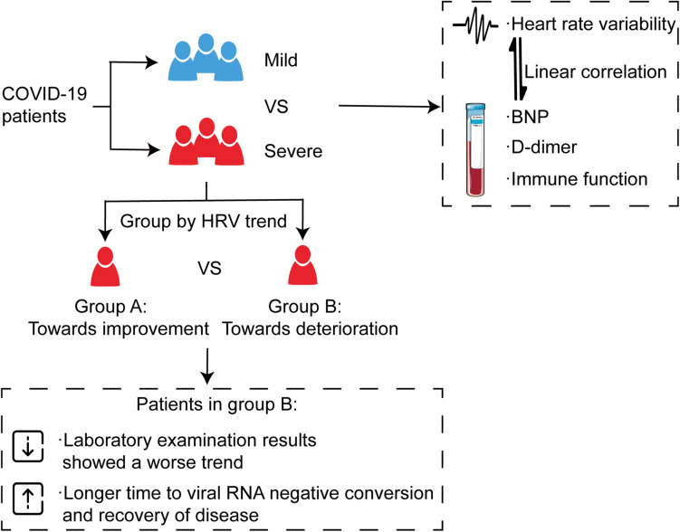
Methods: We included consecutive patients with COVID-19 admitted to the 21st COVID-19 Department of the east campus of Renmin Hospital of Wuhan University from February 6 to March 7, 2020. Clinical data were collected. Heart rate variability (HRV), N-terminal pro-B-type natriuretic peptide (NT-proBNP), D-dimer, and lymphocytes and subsets counts were analysed at two time points: nucleic-acid test positive and negative. Psychological symptoms were assessed after discharge.

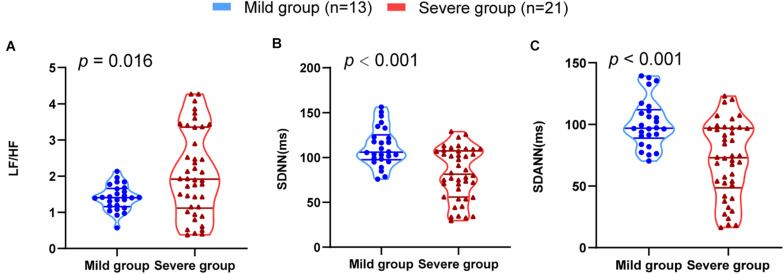
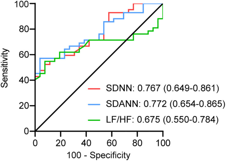
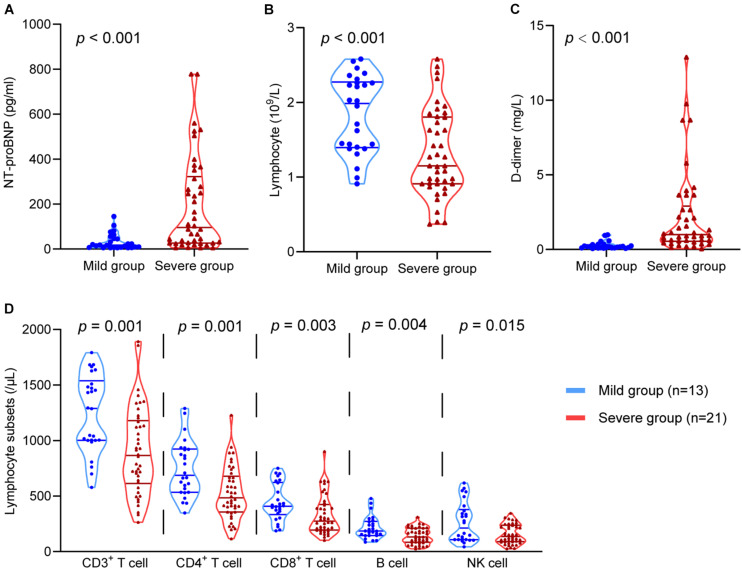
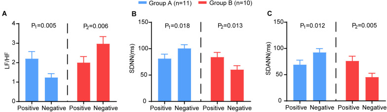
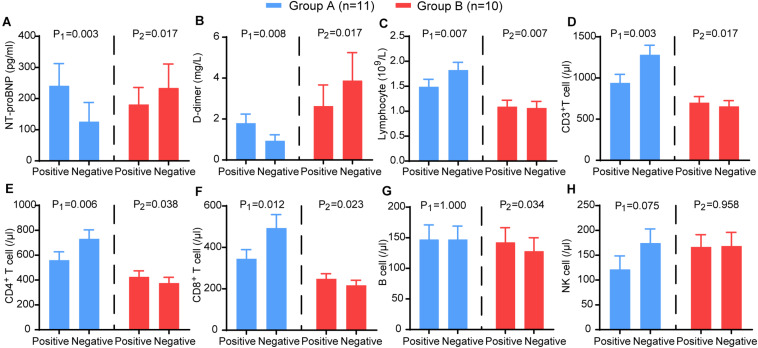
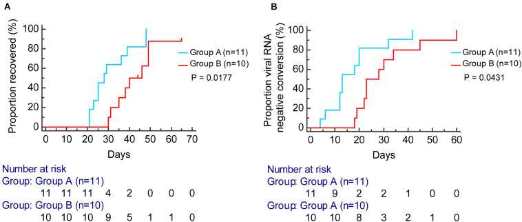
Results: All patients were divided into a mild group (13) and a severe group (21). The latter was further divided into two categories according to the trend of HRV. Severe patients had a significantly lower standard deviation of the RR intervals (SDNN) (P < 0.001), standard deviation of the averages of NN intervals (SDANN) (P < 0.001), and a higher ratio of low- to high-frequency power (LF/HF) (P = 0.016). Linear correlations were shown among SDNN, SDANN, LF/HF, and laboratory indices (P < 0.05). Immune function, D-dimer, and NT-proBNP showed a consistent trend with HRV in severe patients (P < 0.05), and severe patients without improved HRV parameters needed a longer time to clear the virus and recover (P < 0.05).

Conclusion: HRV was associated with the severity of COVID-19. The changing trend of HRV was related to the prognosis, indicating that HRV measurements can be used as a non-invasive predictor for clinical outcome.

Keywords: COVID-19; N-terminal Pro-B-type natriuretic peptide; autonomic nervous system; clinical outcomes; d-dimer; heart rate variability; lymphocyte.


Conflict of interest statement

The authors declare that the research was conducted in the absence of any commercial or financial relationships that could be construed as a potential conflict of interest.

Figures

FIGURE 1
FIGURE 1
HRV measurements in mild and severe patients with COVID-19. (A) LF/HF (B) SDNN (C) SDANN are shown.
FIGURE 2
FIGURE 2
ROC analysis for significant HRV variables. The sensitivity and specificity of SDNN, SDANN, and LF/HF for the severity of COVID-19.
FIGURE 3
FIGURE 3
The comparison of the D-dimer, NT-proBNP, lymphocytes, and lymphocyte subsets in the mild group and the severe group. The comparison of (A) NT-proBNP, (B) lymphocytes, (C) D-dimer, (D) CD3+ T cells, CD4+ T cells, CD8+ T cells, B cells, and NK cells in different groups.
FIGURE 4
FIGURE 4
The correlation between HRV and NT-proBNP, D-dimer, and lymphocytes. (A) LF/HF and NT-proBNP, (B) SDANN and NT-proBNP, (C) SDNN and NT-proBNP, (D) LF/HF and D-dimer, (E) SDANN and D-dimer, (F) SDNN and D-dimer, (G) LF/HF and lymphocytes, (H) SDANN and lymphocytes, (I) SDNN and lymphocytes.
FIGURE 5
FIGURE 5
The comparison of the HRV variables between the two time points (nucleic acid test positive and negative) in group A (blue) and group B (red). The (A) LF/HF, (B) SDNN, (C) SDANN in different groups. P-Values were provided for comparison as follows: P1: Nucleic acid test positive vs nucleic acid test negative for group A. P2: Nucleic acid test positive vs nucleic acid test negative for group B.
FIGURE 6
FIGURE 6
The comparison of the laboratory examination variables between the two time points (nucleic acid test positive and negative) in group A (blue) and group B (red). The (A) NT-proBNP, (B) D-dimer, (C) CD3+ T cells, (D) CD4+ T cells, (E) CD8+ T cells, (F) B cells, (G) NK cells, (H) lymphocytes in different groups. P-values were provided for comparison as follows: P1: Nucleic acid test positive vs nucleic acid test negative for group A. P2: Nucleic acid test positive vs nucleic acid test negative for group B.
FIGURE 7
FIGURE 7
Kaplan–Meier curve analysis for the proportion of viral RNA negative conversion (A) and recovered (B) stratified by group A (blue) and group B (red).
FIGURE 8
FIGURE 8
Alteration of autonomic nervous system is associated with severity and outcomes in patients with COVID-19. All patients were divided into a mild group and a severe group, and the severe group was then further divided into two categories according to the trend of HRV indices, which showed a consistent trend with immune function, D-dimer, and NT-proBNP. The results showed that severe patients without improved HRV parameters needed a longer time to clear the virus and recover.

Similar articles

Cited by

References

    1. Ahmad S., Tejuja A., Newman K. D., Zarychanski R., Seely A. J. (2009). Clinical review: a review and analysis of heart rate variability and the diagnosis and prognosis of infection. Crit. Care 13:232. 10.1186/cc8132 - DOI - PMC - PubMed
    1. Akhmerov A., Marban E. (2020). COVID-19 and the Heart. Circ. Res. 126 1443–1455. 10.1161/CIRCRESAHA.120.317055 - DOI - PMC - PubMed
    1. Barnaby D. P., Fernando S. M., Herry C. L., Scales N. B., Gallagher E. J., Seely A. (2019). Heart rate variability, clinical and laboratory measures to predict future deterioration in patients presenting with sepsis. Shock 51 416–422. 10.1097/SHK.0000000000001192 - DOI - PubMed
    1. Bonaz B., Picq C., Sinniger V., Mayol J. F., Clarençon D. (2013). Vagus nerve stimulation: from epilepsy to the cholinergic anti-inflammatory pathway. Neurogastroenterol. Motil. 25 208–221. 10.1111/nmo.12076 - DOI - PubMed
    1. Bonaz B., Sinniger V., Hoffmann D., Clarençon D., Mathieu N., Dantzer C., et al. (2016). Chronic vagus nerve stimulation in Crohn’s disease: a 6-month follow-up pilot study. Neurogastroenterol. Motil. 28 948–953. 10.1111/nmo.12792 - DOI - PubMed