Review
. 2021 May 18:12:689678.
doi: 10.3389/fendo.2021.689678. eCollection 2021.

Proglucagon-Derived Peptides as Therapeutics

Affiliations
Review

Proglucagon-Derived Peptides as Therapeutics

Ryan A Lafferty et al. Front Endocrinol (Lausanne). .

Abstract

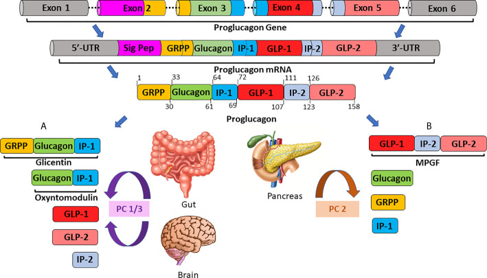
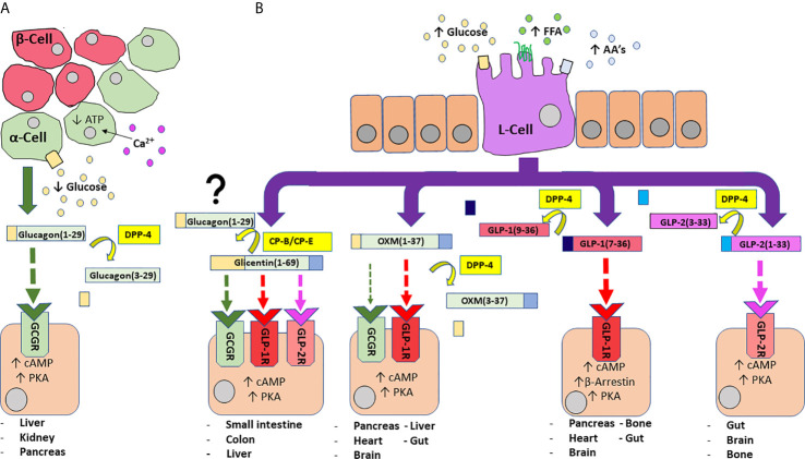
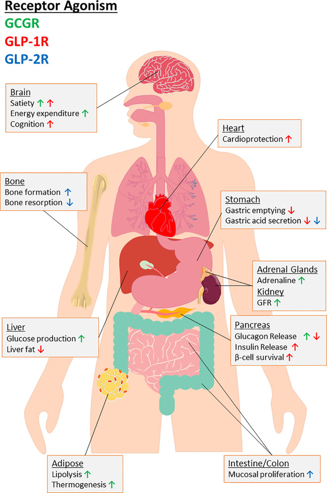
Initially discovered as an impurity in insulin preparations, our understanding of the hyperglycaemic hormone glucagon has evolved markedly over subsequent decades. With description of the precursor proglucagon, we now appreciate that glucagon was just the first proglucagon-derived peptide (PGDP) to be characterised. Other bioactive members of the PGDP family include glucagon-like peptides -1 and -2 (GLP-1 and GLP-2), oxyntomodulin (OXM), glicentin and glicentin-related pancreatic peptide (GRPP), with these being produced via tissue-specific processing of proglucagon by the prohormone convertase (PC) enzymes, PC1/3 and PC2. PGDP peptides exert unique physiological effects that influence metabolism and energy regulation, which has witnessed several of them exploited in the form of long-acting, enzymatically resistant analogues for treatment of various pathologies. As such, intramuscular glucagon is well established in rescue of hypoglycaemia, while GLP-2 analogues are indicated in the management of short bowel syndrome. Furthermore, since approval of the first GLP-1 mimetic for the management of Type 2 diabetes mellitus (T2DM) in 2005, GLP-1 therapeutics have become a mainstay of T2DM management due to multifaceted and sustainable improvements in glycaemia, appetite control and weight loss. More recently, longer-acting PGDP therapeutics have been developed, while newfound benefits on cardioprotection, bone health, renal and liver function and cognition have been uncovered. In the present article, we discuss the physiology of PGDP peptides and their therapeutic applications, with a focus on successful design of analogues including dual and triple PGDP receptor agonists currently in clinical development.

Keywords: GLP-1; GLP-2; diabetes; glucagon; multi-agonist; obesity; oxyntomodulin; proglucagon.


Conflict of interest statement

PF, VG, NI,and FO’H are named on patents filed by Ulster University for the exploitation of incretin-based drugs and other peptide therapeutics. The remaining authors declare that the research was conducted in the absence of any commercial or financial relationships that could be construed as a potential conflict of interest.

Figures

Figure 1
Figure 1
A schematic overview of tissue-specific proglucagon processing in the gut/brain (A) and in the pancreas (B). The proglucagon gene, located on chromosome 2 and comprised of 6 exons, is transcribed to generate proglucagon messenger RNA (mRNA). Proglucagon mRNA is subsequently translated to yield the 158 residue, precursor protein, proglucagon. In enteroendocrine L-cells of the ileum and colon (A) proglucagon is processed by prohormone convertase 1/3 (PC1/3) to generate glicentin, oxyntomodulin, glucagon-like peptides-1 and -2 (GLP-1, GLP-2) and intervening peptide-2 (IP-2). Conversely, in pancreatic alpha-cells (B), post-translational modification by prohormone convertase 2 (PC2) is responsible for the generation of the major proglucagon fragment (MPGF), glucagon, glicentin-related pancreatic polypeptide (GRPP) and intervening peptide-1 (IP-1).
Figure 2
Figure 2
An overview of PGDP actions and secretion from pancreatic alpha-cells (A) and enteroendocrine L-cells (B)). A fall in circulating glucose concentration sees a reduction in intracellular adenosine triphosphate (ATP) levels and resultant closure of ATP-sensitive K+ channels to depolarise the plasma membrane and trigger the influx of Ca2+ ions, the primary stimulus for glucagon release (A). Glucagon is subject to N-terminal dipeptide removal by dipeptidyl-peptidase 4 (DPP-4). Glucagon(1-29) agonises glucagon receptors (GCGR) to evoke protein kinase A (PKA) activation and subsequent mobilisation of cyclic adenosine monophosphate (cAMP). Enteroendocrine L-cells of the distal gut are an open-type cell, rich in chemoreceptors and respond to digestion products of dietary carbohydrate, free fatty acids (FFA) and amino acids (AA’s) to release a number of PGDP’s into circulation (B). Glicentin(1-69) is an agonist for GCGR, GLP-1R and GLP-2R, although with less affinity than their primary hormonal ligands. Additionally, glicentin may serve as a precursor to glucagon in the gut, facilitated enzymatic degradation by enzymes such as carboxypeptidases-B and -E (CP-B, CP-E). Oxyntomodulin (OXM) is a dual agonist for GCGR and GLP-1R, but shows bias towards GLP-1R. It is cleaved by DPP-4 to yield inactive OXM(3-37). Bioactive glucagon-like peptide 1 (GLP-1(7-36)) agonises target GLP-1R to evoke PKA-mediated rises in cAMP, while activation of β-arrestin is also implicated in insulin secretion. DPP-4 cleaved GLP-1(9-36) is inactive. Glucagon-like peptide 2 (GLP-2) agonises target GLP-2R to evoke rises in PKA/cAMP. It is inactivated by DPP-4 to generate GLP-2(3-23). Enzymes are indicated by yellow boxes/arrows. Receptor interactions are indicated by dashed lines, with affinity indicated by increasing thickness of the arrow. Major tissues expressing receptors are also provided.
Figure 3
Figure 3
An overview of the biological consequences for agonism of target receptors of major PGDP’s, namely glucagon receptor (GCGR) and glucagon-like peptide-1 and -2 receptors (GLP-1R, GLP-2R). Organ-specific actions are provided with arrows indicating up or downregulation of specific effects to highlight the therapeutic potential for multiagonism in relation to PGDP’s. As indicated by the key, the colour of arrow indicates the receptor interactions responsible. “GFR” indicates glomerular filtration rate.

Similar articles

Cited by

References

    1. Edkins JS. The Chemical Mechanism of Gastric Secretion. J Physiol (1905) 34:133–44. 10.1113/jphysiol.1906.sp001146 - DOI - PMC - PubMed
    1. Bayliss WM, Starling EH. The Mechanism of Pancreatic Secretion. J Physiol (1902) 28:325–53. 10.1113/jphysiol.1902.sp000920 - DOI - PMC - PubMed
    1. Banting FG, Best CH, Collip JB, Campbell WR, Fletcher AA. Pancreatic Extracts in the Treatment of Diabetes Mellitus. Can Med Assoc J (1922) 12:141–6. - PMC - PubMed
    1. Lawrence RD, Archer N. Zinc Protamine Insulin a Clinical Trial of the New Preparation. Br Med J (1937) 1:487–91. 10.1136/bmj.1.3974.487 - DOI - PMC - PubMed
    1. Bell GI, Pictet RL, Rutter WJ, Cordell B, Tischer E, Goodman HM. Sequence of the Human Insulin Gene. Nature (1980) 284:26–32. 10.1038/284026a0 - DOI - PubMed

Publication types

MeSH terms