. 2021 May 19:12:658373.
doi: 10.3389/fmicb.2021.658373. eCollection 2021.

Understanding How Staphylococcal Autolysin Domains Interact With Polystyrene Surfaces

Affiliations

Understanding How Staphylococcal Autolysin Domains Interact With Polystyrene Surfaces

Radha P Somarathne et al. Front Microbiol. .

Abstract

Biofilms, when formed on medical devices, can cause malfunctions and reduce the efficiency of these devices, thus complicating treatments and serving as a source of infection. The autolysin protein of Staphylococcus epidermidis contributes to its biofilm forming ability, especially on polystyrene surfaces. R2ab and amidase are autolysin protein domains thought to have high affinity to polystyrene surfaces, and they are involved in initial bacterial attachment in S. epidermidis biofilm formation. However, the structural details of R2ab and amidase binding to surfaces are poorly understood. In this study, we have investigated how R2ab and amidase influence biofilm formation on polystyrene surfaces. We have also studied how these proteins interact with polystyrene nanoparticles (PSNPs) using biophysical techniques. Pretreating polystyrene plates with R2ab and amidase domains inhibits biofilm growth relative to a control protein, indicating that these domains bind tightly to polystyrene surfaces and can block bacterial attachment. Correspondingly, we find that both domains interact strongly with anionic, carboxylate-functionalized as well as neutral, non-functionalized PSNPs, suggesting a similar binding interaction for nanoparticles and macroscopic surfaces. Both anionic and neutral PSNPs induce changes to the secondary structure of both R2ab and amidase as monitored by circular dichroism (CD) spectroscopy. These changes are very similar, though not identical, for both types of PSNPs, suggesting that carboxylate functionalization is only a small perturbation for R2ab and amidase binding. This structural change is also seen in limited proteolysis experiments, which exhibit substantial differences for both proteins when in the presence of carboxylate PSNPs. Overall, our results demonstrate that the R2ab and amidase domains strongly favor adsorption to polystyrene surfaces, and that surface adsorption destabilizes the secondary structure of these domains. Bacterial attachment to polystyrene surfaces during the initial phases of biofilm formation, therefore, may be mediated by aromatic residues, since these residues are known to drive adsorption to PSNPs. Together, these experiments can be used to develop new strategies for biofilm eradication, ensuring the proper long-lived functioning of medical devices.

Keywords: autolysin proteins; biofilms; medical implants; polystyrene nanoparticles; surface chemistry.


Conflict of interest statement

The authors declare that the research was conducted in the absence of any commercial or financial relationships that could be construed as a potential conflict of interest.

Figures

FIGURE 1
FIGURE 1
The effect of protein treatment on biofilm formation. Wells were pre-coated with either no protein (control), amidase, R2ab, or BSA, followed by growth conditions favoring biofilm formation, which was monitored by crystal violet staining and measurement of absorbance at 570 nm. The BSA-coated plate showed only a slight reduction in biofilm growth relative to no treatment, but plates treated with amidase and R2ab domains show a much larger reduction. A typical row from a stained, 96-well plate is shown above each corresponding column to display the results of crystal violet staining. The error bars for each data point represent the standard error of the mean for N = 8 samples. A one-way ANOVA test [F(3,28) = 217.2, p < 0.0001] with Tukey’s post hoc analysis was performed to determine the levels of significance (**p < 0.005, ****p < 0.0001).
FIGURE 2
FIGURE 2
Representative microbial growth curves in the presence of R2ab (A) and amidase (C). Growth curves are shown for S. epidermidis when the media was mixed with (A,B) R2ab or (C,D) amidase. Concentrations added for each protein domain are 0 μM (red), 2 μM (orange), 4 μM (yellow), 6 μM (green), 8 μM (blue), and 10 μM (purple). The black curve shows a control with no bacteria added. Panels (B,D) show the average and standard deviation of OD600 at 18 h for R2ab and amidase, respectively, for the different treatments (N = 8). A one-way ANOVA test [(B): F(5, 42) = 120.4, p < 0.0001 and (D): F(5, 41) = 77.51, p < 0.0001] with Tukey’s post hoc analysis was performed to determine the levels of significance between groups (n.s. is not significant; **p < 0.005 and ****p < 0.0001).
FIGURE 3
FIGURE 3
Zeta potential changes when carboxylate functionalized PSNPs are titrated with autolysin domains. The zeta potential of 20 nm (nominal diameter) carboxylate-functionalized polystyrene nanospheres increases when R2ab or amidase domains are added to solution, but very little change is seen for BSA. Error bars represent the standard deviation for N = 3 experiments, and lines connecting each point are added as a guide to the eye.
FIGURE 4
FIGURE 4
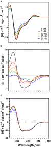
Circular dichroism spectra for the of autolysin domains and a negative control in the presence of carboxylate functionalized PSNPs. R2ab (A) and amidase (B) exhibit changes in their CD spectra when nanoparticle concentrations are increased. In the absence of nanoparticles (black curve), both domains exhibit a well-defined secondary structure. As nanoparticles are increased to 5 nM (red), 10 nM (orange), 15 nM (green), and 20 nM (blue), secondary structure changes become evident. The same changes are not observed for BSA (C), whose spectrum remains fairly constant as PSNP concentration is altered.
FIGURE 5
FIGURE 5
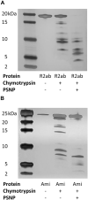
Limited proteolysis of autolysin domains. Representative silver-stained products of chymotrypsin cleavage of R2ab (A) and amidase [Ami, (B)] domains in the presence and absence of carboxylate functionalized PSNPs. The leftmost lane contains markers, and molecular weights are labeled. Limited proteolysis is more complete in the presence of PSNPs for both proteins, suggesting partial unfolding in the presence of nanoparticles.

References

    1. Andrade J. D., Hlady V. (1986). “Protein adsorption and materials biocompatibility: a tutorial review and suggested hypotheses,” in Biopolymers/Non-Exclusion HPLC, ed. Dusek K. (Berlin: Spinger-Verlag; ), 10.1007/3-540-16422-7_6 - DOI
    1. Baier G., Costa C., Zeller A., Baumann D., Sayer C., Araujo P. H. H., et al. (2011). BSA adsorption on differently charged polystyrene nanoparticles using isothermal titration calorimetry and the influence on cellular uptake. Macromol. Biosci. 11 628–638. 10.1002/mabi.201000395 - DOI - PubMed
    1. Billings N., Millan M. R., Caldara M., Rusconi R., Tarasova Y., Stocker R., et al. (2013). The extracellular matrix component Psl provides fast-acting antibiotic defense in Pseudomonas aeruginosa biofilms. PLoS Pathogens 9:e1003526. 10.1371/journal.ppat.1003526 - DOI - PMC - PubMed
    1. Brownridge P., Beynon R. J. (2011). The importance of the digest: proteolysis and absolute quantification in proteomics. Methods 54 351–360. 10.1016/j.ymeth.2011.05.005 - DOI - PubMed
    1. Büttner F. M., Zoll S., Nega M., Götz F., Stehle T. (2014). Structure-function analysis of Staphylococcus aureus amidase reveals the determinants of peptidoglycan recognition and cleavage. J. Biol. Chem. 289 11083–11094. 10.1074/jbc.M114.557306 - DOI - PMC - PubMed

LinkOut - more resources