. 2021 May 18:12:609543.
doi: 10.3389/fimmu.2021.609543. eCollection 2021.

Analysis of Porcine RIG-I Like Receptors Revealed the Positive Regulation of RIG-I and MDA5 by LGP2

Affiliations

Analysis of Porcine RIG-I Like Receptors Revealed the Positive Regulation of RIG-I and MDA5 by LGP2

Shuangjie Li et al. Front Immunol. .

Abstract

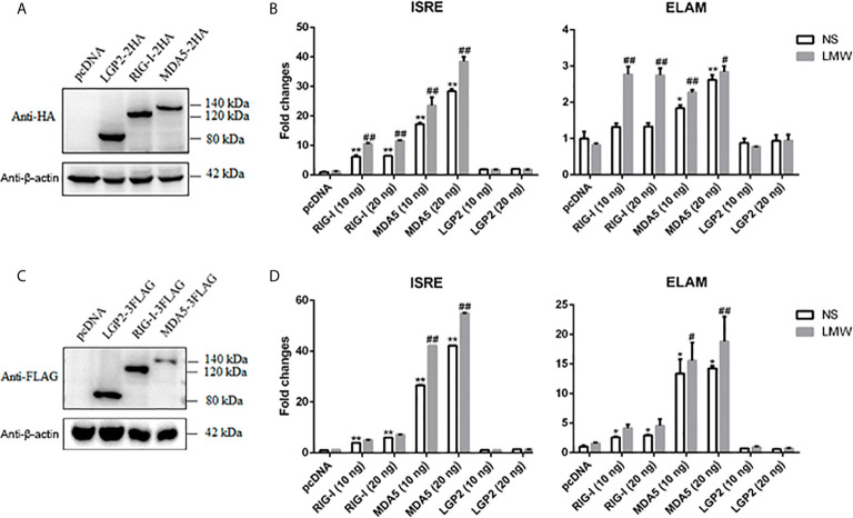
The RLRs play critical roles in sensing and fighting viral infections especially RNA virus infections. Despite the extensive studies on RLRs in humans and mice, there is a lack of systemic investigation of livestock animal RLRs. In this study, we characterized the porcine RLR members RIG-I, MDA5 and LGP2. Compared with their human counterparts, porcine RIG-I and MDA5 exhibited similar signaling activity to distinct dsRNA and viruses, via similar and cooperative recognitions. Porcine LGP2, without signaling activity, was found to positively regulate porcine RIG-I and MDA5 in transfected porcine alveolar macrophages (PAMs), gene knockout PAMs and PK-15 cells. Mechanistically, LGP2 interacts with RIG-I and MDA5 upon cell activation, and promotes the binding of dsRNA ligand by MDA5 as well as RIG-I. Accordingly, porcine LGP2 exerted broad antiviral functions. Intriguingly, we found that porcine LGP2 mutants with defects in ATPase and/or dsRNA binding present constitutive activity which are likely through RIG-I and MDA5. Our work provided significant insights into porcine innate immunity, species specificity and immune biology.

Keywords: LGP2; RIG-like receptors (RLRs); porcine; positive regulation; species specificity.


Conflict of interest statement

The authors declare that the research was conducted in the absence of any commercial or financial relationships that could be construed as a potential conflict of interest.

Figures

Figure 1
Figure 1
The expression of porcine LGP2, RIG-I, MDA5 and their activity in promoter assays. (A) The pcDNA-pLGP2-HA, pcDNA-pRIG-I-HA, pcDNA-pMDA5-HA and vector pcDNA (0.5 μg each) were transfected into 293T cells in 24-well plate (3×10⁵ cells/well) for 24 h using Lipofectamine 2000. The cells samples were detected by Western-blotting with anti-HA mAb. (B) 293T cells grown in 96-well plates (1×10⁴ cells/well) were transfected with 10 ng or 20 ng pcDNA-pLGP2-HA, pcDNA-pRIG-I-HA, pcDNA-pMDA5-HA, plus ISRE-Fluc/ELAM-Fluc (10 ng) and Rluc (0.2 ng), which were normalized to 50 ng/well by vector pcDNA. Twenty-four hours post transfection, the cells were transfected or not with LMW poly I:C (1 μg/ml) for 8 h. The luciferase activities were measured with Double-Luciferase Reporter Assay. (C) The pcDNA-pLGP2-FLAG, pcDNA-pRIG-I-FLAG, pcDNA-pMDA5-FLAG and pcDNA were transfected as in (A) and detected with anti-FLAG mAb. (D) Promoter activation by pcDNA-pLGP2-FLAG, pcDNA-pRIG-I-FLAG, pcDNA-pMDA5-FLAG were detected as in (B). *p < 0.05, **p < 0.01 vs pcDNA controls. # p < 0.05, ## p < 0.01 vs non-stimulated (NS) controls.
Figure 2
Figure 2
The signal activity of pLGP2, pRIG-I and pMDA5 before and after poly I:C transfection and virus infection. (A) 293T cells grown in 24-well plates (3×10⁵ cells/well) were transfected with HA tagged pLGP2, pRIG-I, pMDA5 or pcDNA (0.5 μg each) using Lipofectamine 2000. Twenty-four hours post transfection, the cells were transfected with LMW poly I:C or HMW poly I:C (1 μg/ml) for 8h stimulation. The harvested cells were analyzed by RT-qPCR for downstream gene expressions as indicated. (B) 293T cells in 24-well plates (3×10⁵ cells/well) were transfected as in (A) and infected with 0.01 MOI EMCV or VSV for 8 h, and analyzed by RT-qPCR. *p < 0.05, **p < 0.01 vs pcDNA controls. # p < 0.05, ## p < 0.01 vs NS controls.
Figure 3
Figure 3
LGP2 promoted the expression of downstream genes of RIG-I and MDA5 in PAMs after LMW poly I:C transfection and VSV infection. (A) PAMs grown in 24-well plates (3×10⁵ cells/well) were transfected with LGP2 in different amounts (0, 0.25, 0.5, 1 μg) using Lipofectamine 3000. Twenty-four hours post transfection, the cells were stimulated with LMW poly I:C transfection (1 μg/ml) for 8h. Then the cells were analyzed by RT-qPCR for downstream gene expressions as indicated. (B) PAMs grown in 24-well plates were transfected as in (A). The cells were infected with 0.01 MOI VSV for 8h and analyzed by RT-qPCR. *p < 0.05, **p < 0.01 vs mock transfection and NS controls. # p < 0.05, ## p < 0.01 vs mock transfection and stimulated controls.
Figure 4
Figure 4
Expressions of downstream genes in CRISPR KO stable cell lines upon stimulations. (A) The LGP2, RIG-I and MDA5 stable KO PAMs plus CRISPR control PAMs grown in 24-well plates (3×10⁵ cells/well) were transfected with LMW poly I:C (1 μg/ml). Eight hours post transfection, the cells were analyzed by RT-qPCR for downstream gene expressions. (B) The LGP2, RIG-I, MDA5 stable KO and control PAMs were infected with VSV at MOI of 0.01 for 8h and analyzed as in (A). (C) The LGP2, RIG-I and MDA5 KO PK15 cells plus CRISPR control PK15 cells grown in 24-well plates (3×10⁵ cells/well) were transfected and analyzed as in (A). (D, E) The LGP2, RIG-I, MDA5 stable KO PK15 cells were infected with VSV (D) or EMCV (E) at MOI of 0.01 and analyzed as in (A). **p < 0.01 vs CRISPR NS controls. # p < 0.05, ## p < 0.01 vs CRISPR stimulated controls.
Figure 5
Figure 5
Expressions of cellular genes and viral replications in LGP2, RIG-I and MDA5 homozygous KO PAM cell clones. (A) The LGP2-/-, RIG-I-/- and MDA5-/- PAMs plus CRISPR control PAMs grown in 12-well plates (5×10⁵ cells/well) were transfected with LMW poly I:C or HMW poly I:C (1 μg/ml) for 8h. The expression of downstream cellular genes were analyzed by RT-qPCR. (B) The LGP2-/-, RIG-I-/- and MDA5-/- PAMs plus CRISPR control PAMs grown in 12-well plates (5×10⁵ cells/well) were infected with EMCV at 0.01 MOI for 8 h. The cells were detected by Western-blotting with the indicated antibodies. (C, D) The LGP2-/-, RIG-I-/- and MDA5-/- PAMs plus CRISPR control PAMs in 12-well plates (5×10⁵ cells/well) were infected with various viruses at MOI of 0.01 for 8-12 h and analyzed by RT-qPCR for the expressions of downstream cellular genes (C) and viral genes (up panel, D). The supernatants from VSV and HSV1 infected PAMs were subjected to infection of Vero cells and the viral plaques were visualized at 30 h and 72 h post infection, respectively. The countable plaque numbers are labeled in red color below the wells of cell plates (low panel, D). (E, F) MDA5-/- PAMs (E) and RIG-I-/- PAMs (F) in 24-well plates were transfected different amounts of LGP2 and stimulated with 0.01 MOI VSV for 8 h. The downstream IFNβ and ISG56 were analyzed by RT-qPCR. **p < 0.01 vs NS controls. # p < 0.05, ## p < 0.01 vs CRISPR controls in (A–D), or pcDNA VSV controls in (E, F).
Figure 6
Figure 6
The antiviral effects of ectopic porcine LGP2, RIG-I and MDA5. (A, B) PAMs were infected with swine influenza virus H9N2 at MOI of 0.01 for different hours and the viral protein NS1 was detected by Western-blotting with anti-NS1 mAb (left panel, A). PAMs grown in 12-well plates (5×10⁵ cells/well) were transfected with LGP2, RIG-I and MDA5 (1 μg each) using TransIT-LT1 Transfection Reagent. The cells were infected with 0.01 MOI H9N2 for further 63 h and analyzed by Western-blotting for NS1 protein expression (right panel, A) and RT-qPCR for viral HA and M gene expressions (B). (C) IPEC-J2 cells grown in 12-well plates (5×10⁵ cells/well) were transfected as in (A) and infected with 0.01 MOI PEDV (YC2014 strain) for 24 h. The cells were analyzed by RT-qPCR for viral M gene expression (left). The cell supernatants were examined by TCID50 assay (middle) and plaque assay (right), which were performed with Vero cells for 6 day infection and the countable plaque numbers are labeled in red color below the wells of plate. (D, E) Marc-145 cells were infected with 0.01 MOI PRRSV (JXA1-R strain) for different hours and the virus protein N was detected by Western-blotting with anti-N mAb (left panel, D). Marc-145 cells grown in 12-well plates (5×10⁵ cells/well) were transfected as in (A), and the cells were infected with 0.01 MOI JXA1-R for 36h and analyzed by Western-blotting for N protein expression (right panel, D) and RT-qPCR for viral N gene (left, E). The cell supernatants were examined by TCID50 assay (middle, E) and plaque assay (right, E), which were performed with Marc-145 cells for 6 day infection and the countable plaque numbers are labeled in red color below the wells of plate *p < 0.05, **p < 0.01 vs pcDNA controls.
Figure 7
Figure 7
Porcine LGP2 interacts RIG-I and MDA5 upon activation and promotes their RNA binding. (A) pLGP2-FLAG (1 μg) and pRIG-I-HA (1.5 μg) were co-transfected into 293T cells in 6-well plate (8×10⁵ cells/well) for 24 h, then infected with or without 0.01 MOI EMCV for 8 h. The cells lysates were immunoprecipitated with anti-FLAG mAb and subjected to Western-blotting using the indicated antibodies. (B) pLGP2-FLAG and pMDA5-HA were co-transfected into 293T, and the transfected cells were treated and processed as in (A). (C) pMDA5-FLAG and pRIG-I-HA were co-transfected into 293T cells, and the transfected cells were treated and processed as in (A). (D) PAMs grown on 15-mm glass bottom cell culture dish (5×10⁵ cells) were co-transfected with pLGP2-HA (0.75 μg) and pRIG-I-FLAG (0.75 μg) for 24 h, then infected with or without EMCV for 8 h. The cells were examined for co-localization by con-focal fluorescence microscopy. (E) pLGP2-HA and pMDA5-FLAG were co-transfected and the transfected PAMs were treated and examined for co-localization as in (D). (F) pRIG-HA and pMDA5-FLAG were co-transfected, and transfected PAMs were treated and examined for co-localization as in (D). (G) The purified pLGP2-HA, pLGP2-FLAG, pRIG-HA and pMDA5-FLAG were analyzed by SDS-PAGE. (H) pLGP2-HA, pRIG-I-HA and pMDA5-FLAG were incubated in tubes alone or together as indicated, then 1μg dsRNA poly (I:C) HMW Biotin and 30 μL streptavidin agarose were added for binding. The dsRNA bound proteins were analyzed by Western-blotting.
Figure 8
Figure 8
Expression of pLGP2 mutants and their positive regulation on pRIG-I and pMDA5. (A) The pLGP2 protein structure and domains predicted by UniProt (https://www.uniprot.org). (B) The protein expressions of pLGP2 domains, deletion mutants and point mutants in transfected 293T cells. (C) PAMs grown in 12-well plates (5×10⁵ cells) were transfected with pLGP2 mutants (1 μg each) using TransIT-LT1 Transfection Reagent for 24 h. The cells were analyzed by RT-PCR for the expressions of downstream genes. (D) PAMs grown in 96-well plates (1×10⁴ cells/well) were transfected with pLGP2 and its mutants (20 ng each) plus ISRE-Fluc/ELAM-Fluc (10 ng) and Rluc (0.2 ng), using the TransIT-LT1 Transfection Reagent. Twenty-four hours post transfection, the luciferase activities were measured. (E) PAMs grown in 12-well plates (5×10⁵ cells) were transfected as in (C), then infected with 0.01 MOI VSV-GFP for 12 h. The cells were analyzed by Western-blotting with anti-GFP mAb, and the GFP densitometric values after normalization by actin were shown below. *p < 0.05, **p < 0.01 vs pcDNA controls.
Figure 9
Figure 9
The porcine LGP2 mutant activity in RIG-/- and MDA5-/- PAMs and the antiviral activity. (A) RIG-/-, MDA5-/- and CRISPR control PAMs in 12-well plates (5×10⁵ cells) were transfected with pLGP2 and its mutants (0.75 μg each). Twenty-four hours later, the cells were analyzed by RT-qPCR for the downstream IFNβ gene transcriptions. **p < 0.01 vs pcDNA controls. (B, C) LGP2-/- PAMs were transfected with 0.75 μg pLGP2, its constitutive MIII mutant or pcDNA control (C) for 24 h. PAMs transfected with pcDNA was used as a control (PAM C). The transfected cells were infected with 0.01 MOI VSV, SeV and HSV-1 for 8 h, and analyzed by RT-qPCR for virus replications (B), as well as for cellular downstream IFNβ and ISG56 expressions (C). *p < 0.05, **p < 0.01 vs PAM C or N. I # p < 0.05, ## p < 0.01 vs LGP2-/- PAM C.
Figure 10
Figure 10
Schematic diagram of porcine RIG-I and MDA5 signaling and the regulation by LGP2. Porcine RIG-I and MDA5 similarly recognize dsRNA and sense viruses regardless of the lengths of dsRNA and the affinity of binding to dsRNA, to trigger downstream signaling and gene transcription. The porcine LGP2, binding with dsRNA in a similar way to MDA5, is able to promote the dsRNA binding by both MDA5 and RIG-I, and thus downstream signaling. Intriguingly, during the positive regulation by LGP2, the ATPase hydrolysis and dsRNA binding activity seem dispensable. The solid lines refer to confirmed interactions in this study, whereas the dashed lines are based on previous knowledge.

Similar articles

Cited by

References

    1. Takeuchi O, Akira S. Pattern Recognition Receptors and Inflammation. Cell (2010) 140:805–20. 10.1016/j.cell.2010.01.022 - DOI - PubMed
    1. Cui J, Chen Y, Wang HY, Wang RF. Mechanisms and Pathways of Innate Immune Activation and Regulation in Health and Cancer. Hum Vaccines Immunotherapeut (2014) 10:3270–85. 10.4161/21645515.2014.979640 - DOI - PMC - PubMed
    1. Zou J, Chang M, Nie P, Secombes CJ. Origin and Evolution of the RIG-I Like RNA Helicase Gene Family. BMC Evolutionary Biol (2009) 9:85. 10.1186/1471-2148-9-85 - DOI - PMC - PubMed
    1. Yoneyama M, Kikuchi M, Natsukawa T, Shinobu N, Imaizumi T, Miyagishi M, et al. . The RNA Helicase RIG-I has an Essential Function in Double-Stranded Rna-Induced Innate Antiviral Responses. Nat Immunol (2004) 5:730–7. 10.1038/ni1087 - DOI - PubMed
    1. Andrejeva J, Childs KS, Young DF, Carlos TS, Stock N, Goodbourn S, et al. . The V Proteins of Paramyxoviruses Bind the IFN-inducible Rna Helicase, mda-5, and Inhibit its Activation of the IFN-beta Promoter. Proc Natl Acad Sci USA (2004) 101:17264–9. 10.1073/pnas.0407639101 - DOI - PMC - PubMed

Publication types

LinkOut - more resources