

A Profound Membrane Reorganization Defines Susceptibility of Plasmodium falciparum Infected Red Blood Cells to Lysis by Granulysin and Perforin
- PMID: 34093532
- PMCID: PMC8170093
- DOI: 10.3389/fimmu.2021.643746
A Profound Membrane Reorganization Defines Susceptibility of Plasmodium falciparum Infected Red Blood Cells to Lysis by Granulysin and Perforin
Abstract
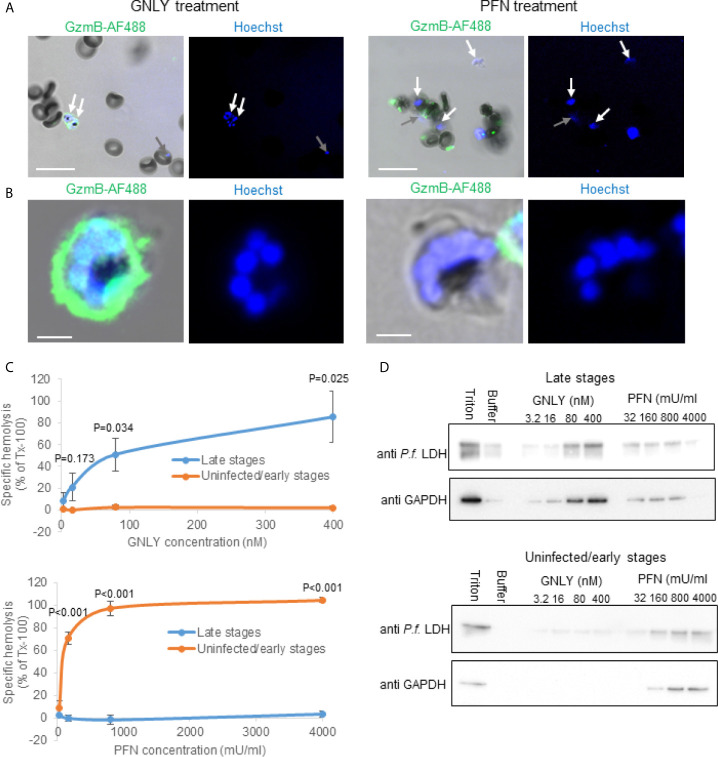
Malaria remains one of the most serious health problems in developing countries. The causative agent of malaria, Plasmodium spp., have a complex life cycle involving multiple developmental stages as well as different morphological, biochemical and metabolic requirements. We recently found that γδ T cells control parasite growth using pore-forming proteins to deliver their cytotoxic proteases, the granzymes, into blood residing parasites. Here, we follow up on the molecular mechanisms of parasite growth inhibition by human pore-forming proteins. We confirm that Plasmodium falciparum infection efficiently depletes the red blood cells of cholesterol, which renders the parasite surrounding membranes susceptible to lysis by prokaryotic membrane disrupting proteins, such as lymphocytic granulysin or the human cathelicidin LL-37. Interestingly, not the cholesterol depletion but rather the simultaneous exposure of phosphatidylserine, a negatively charged phospholipid, triggers resistance of late stage parasitized red blood cells towards the eukaryotic pore forming protein perforin. Overall, by revealing the molecular events we establish here a pathogen-host interaction that involves host cell membrane remodeling that defines the susceptibility towards cytolytic molecules.
Keywords: blood-stage malaria; cholesterol; granulysin; perforin; phosphatidylserine (PS); plasma membrane; pore forming proteins (PFPs).
Copyright © 2021 Hernández-Castañeda, Lavergne, Casanova, Nydegger, Merten, Subramanian, Matthey, Lannes, Mantel and Walch.
Conflict of interest statement
The authors declare that the research was conducted in the absence of any commercial or financial relationships that could be construed as a potential conflict of interest.
Figures


Similar articles
-
Control of Plasmodium falciparum erythrocytic cycle: γδ T cells target the red blood cell-invasive merozoites.Blood. 2011 Dec 22;118(26):6952-62. doi: 10.1182/blood-2011-08-376111. Epub 2011 Nov 1. Blood. 2011. PMID: 22045985
-
γδ T Cells Kill Plasmodium falciparum in a Granzyme- and Granulysin-Dependent Mechanism during the Late Blood Stage.J Immunol. 2020 Apr 1;204(7):1798-1809. doi: 10.4049/jimmunol.1900725. Epub 2020 Feb 17. J Immunol. 2020. PMID: 32066596 Free PMC article.
-
Human gamma delta T cells that inhibit the in vitro growth of the asexual blood stages of the Plasmodium falciparum parasite express cytolytic and proinflammatory molecules.Scand J Immunol. 1999 Dec;50(6):642-50. doi: 10.1046/j.1365-3083.1999.00647.x. Scand J Immunol. 1999. PMID: 10607313
-
Human Vγ9Vδ2 T Lymphocytes in the Immune Response to P. falciparum Infection.Front Immunol. 2018 Nov 27;9:2760. doi: 10.3389/fimmu.2018.02760. eCollection 2018. Front Immunol. 2018. PMID: 30538708 Free PMC article. Review.
-
Is there a role for gamma delta T cells in malaria?Immunol Today. 1992 Aug;13(8):298-300. doi: 10.1016/0167-5699(92)90041-5. Immunol Today. 1992. PMID: 1387316 Review.
Cited by
-
The role of cholesterol in invasion and growth of malaria parasites.Front Cell Infect Microbiol. 2022 Sep 16;12:984049. doi: 10.3389/fcimb.2022.984049. eCollection 2022. Front Cell Infect Microbiol. 2022. PMID: 36189362 Free PMC article. Review.
-
Oxidative and Non-Oxidative Antimicrobial Activities of the Granzymes.Front Immunol. 2021 Oct 11;12:750512. doi: 10.3389/fimmu.2021.750512. eCollection 2021. Front Immunol. 2021. PMID: 34707614 Free PMC article. Review.
-
Antimalarial activity of cecropin antimicrobial peptides derived from Anopheles mosquitoes.Antimicrob Agents Chemother. 2024 Jul 9;68(7):e0031124. doi: 10.1128/aac.00311-24. Epub 2024 Jun 14. Antimicrob Agents Chemother. 2024. PMID: 38874346 Free PMC article.
-
Role of TAM Receptors in Antimalarial Humoral Immune Response.Pathogens. 2024 Apr 2;13(4):298. doi: 10.3390/pathogens13040298. Pathogens. 2024. PMID: 38668253 Free PMC article. Review.
-
Escaping Death: How Cancer Cells and Infected Cells Resist Cell-Mediated Cytotoxicity.Front Immunol. 2022 Mar 23;13:867098. doi: 10.3389/fimmu.2022.867098. eCollection 2022. Front Immunol. 2022. PMID: 35401556 Free PMC article. Review.
References
-
- WHO . (2019). Malaria.
Publication types
MeSH terms
Substances
LinkOut - more resources
Full Text Sources
Molecular Biology Databases
Miscellaneous