. 2021 May 19:12:662901.
doi: 10.3389/fimmu.2021.662901. eCollection 2021.

Effects of Peptide-Induced Immune Tolerance on Murine Lupus

Affiliations

Effects of Peptide-Induced Immune Tolerance on Murine Lupus

Ram P Singh et al. Front Immunol. .

Abstract

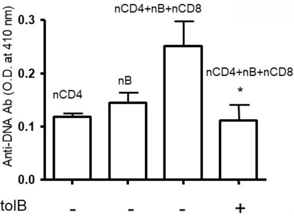
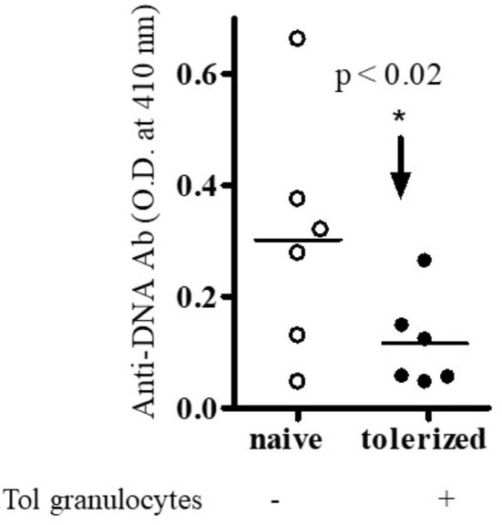
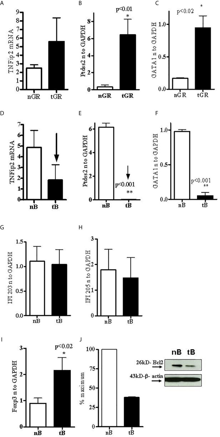
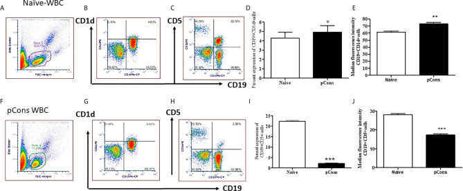
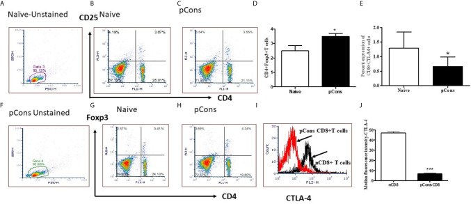
The regulation of autoimmunity and the molecular mechanisms by which different immune cells, including T cells, polymorphonuclear leukocytes (PMN-granulocytes), and B cells suppress autoimmune diseases is complex. We have shown previously that BWF1 lupus mice are protected from autoimmunity after i.v. injection or oral administration of tolerogenic doses of pCons, an artificial synthetic peptide based on sequences containing MHC class I and MHC class II determinants in the VH region of a J558-encoded BWF1 anti-DNA Ab. Several T cell subsets can transfer this tolerance. In this study, we determined the potential roles of granulocytes, B cells and regulatory T cells altered by pCons treatment in the BWF1 (NZB/NZW) mouse model of lupus. Immunophenotyping studies indicated that pCons treatment of BWF1 mice significantly increased CD4+FoxP3+ T cells, reduced the percent of B cells expressing CD19+CD5+ but increased the percent of CD19+CD1d+ regulatory B cells and increased the ability of the whole B cell population to suppress IgG anti-DNA production in vitro. pCons treatment significantly decreased the expression of CTLA-4 (cytotoxic T-lymphocyte-associated protein-4) in CD8+ T cells. In addition, peptide administration modified granulocytes so they became suppressive. We co-cultured sorted naïve B cells from mice making anti-DNA Ab (supported by addition of sorted naive CD4+ and CD8+ T cells from young auto-antibody-negative BWF1 mice) with sorted B cells or granulocytes from tolerized mice. Both tolerized granulocytes and tolerized B cells significantly suppressed the production of anti-DNA in vitro. In granulocytes from tolerized mice compared to saline-treated littermate controls, real-time PCR analysis indicated that expression of interferon-induced TNFAIP2 increased more than 2-fold while Ptdss2 and GATA1 mRNA were up-regulated more than 10-fold. In contrast, expression of these genes was significantly down-regulated in tolerized B cells. Further, another IFN-induced protein, Bcl2, was reduced in tolerized B cells as determined by Western blot analyses. In contrast, expression of FoxP3 was significantly increased in tolerized B cells. Together, these data suggest that B cells and granulocytes are altered toward suppressive functions by in vivo tolerization of BWF1 mice with pCons and it is possible these cell types participate in the clinical benefits seen in vivo.

Keywords: Anti-DNA Ab; granulocytes; immune tolerance and regulation; pConsensus peptide (pCons); polymorphonuclear cells (PMNs); regulatory B cells; systemic lupus erythematosus.


Conflict of interest statement

BHH and RPS have a patent through the University of California, Los Angeles for the use of pCons as an immune modulator in systemic lupus erythematosus. The remaining author declares that the research was conducted in the absence of any commercial or financial relationships that could be construed as a potential conflict of interest.

Figures

Figure 1
Figure 1
Anti-DNA Ab was significantly decreased in the presence of tolerized B cells. Naïve CD4+ T cells, CD8+ T cells and CD45R/B220+B cells were isolated from BWF1 mice spleen cells using microbeads from Miltenyi Biotech (Auburn, CA, USA). Cells were cultured in RPMI 1640 supplemented with L-glutamine (2 mM), penicillin (100 units/ml), streptomycin (0.1 mg/ml), 2-mercaptoethanol (Gibco) and 10% fetal bovine serum (FBS). Cells were co-cultured in the presence of tolerized B cells (1x10⁶ cells). Different immune cell subsets (naïve B cells, 1x10⁵ cells from old nephritic mice; CD4+CD25- T cells, 1x10⁶; naïve CD8+ T cells, 1x10⁵) were isolated from splenocytes and cultured with tolerized B cells (1x10⁶ cells). After the 72-96 hours range, culture supernatants were obtained. Anti-DNA Ab levels were measured from culture supernatants by ELISA. *p < 0.05.
Figure 2
Figure 2
Anti-DNA Ab was significantly decreased in the presence of tolerized PMNs-granulocytes. Different immune cell subsets (naïve B cells, 1x 10⁵ cells from old nephritic mice; CD4+CD25- T cells, 1x10⁶; naïve CD8+ T cells, 1x10⁵) were isolated and cultured with tolerized granulocytes (GR, 1x10⁶). Cell subsets were isolated from total spleen cells of BWF1 mice. Cells were cultured in RPMI 1640 supplemented with L-glutamine (2 mM), penicillin (100 units/ml), streptomycin (0.1 mg/ml), 2-mercaptoethanol (Gibco) and 10% fetal bovine serum (FBS). After the 72- 96 hours range, culture supernatants were obtained. Anti-DNA Ab levels were measured from culture supernatants by ELISA. *p < 0.05.
Figure 3
Figure 3
Tolerized B cells have reduced IFNs gene mRNA and Bcl2 protein level and increased FoxP3 mRNA expression. RNA was isolated from naïve and tolerized B cells and granulocytes. Real time PCR was performed with 100 ng of RNA with gene specific primers and probes. Data was normalized with GAPDH mRNA levels. *p < 0.05. TNFAIP2, Ptdss2, and GATA1 mRNA was increased (A–C) in tolerized granulocytes (GR) but reduced in tolerized B cells (D–F). IFI203 and IFI205 was decreased in tolerized B cells (G, H). (I) FoxP3 expression was increased in tolerized B cells. (J) Quantification of Western blot analysis of Bcl2 protein levels in cell lysates from naïve and tolerized sorted B (CD45R/B220) cells.
Figure 4
Figure 4
pCons treatment modified the cell surface expression markers for regulatory B cells. Female 35-wk old BWF1 mice were treated with pCons (1 mg i.v.). After 3 days, blood was obtained, RBC lysed, and cells were stained with CD19, CD1d and CD5 antibodies and FACS performed. Representative live cell gating strategy (A, F) and FACS analysis of CD1d (B, G) and CD5 (C, H) expression levels from representative naïve (A–C) and pCons treated (F–H) mice. Percent expression of CD19+ CD1d+ cells in naïve vs pCons treated mice (D). Percent expression of CD19+CD5+cells in naïve vs pCons treated mice (I). Quantification of Median Fluorescent Intensity of naïve and pCons-treated mice for CD1d (E) (from B and G Gate 3 and 4) and CD5 (J) (from C and H Gate 3 and 4). CD1d increased and CD5 cells decreased with pCons treatment. *p < 0.05, **p < 0.001, ***p < 0.0001.
Figure 5
Figure 5
pCons treatment increased CD4+FoxP3+ regulatory T cells and significantly reduced Median Fluorescence Intensity of CTLA-4 (Cytotoxic T-lymphocyte-Associated Proten-4) in CD8+ T cells of BWF1 lupus mice. Female 12-20 wk-old BWF1 mice were treated with pCons (1mg i.v.). After 1-2 weeks, splenocytes were obtained, RBC lysed, and cells were stained with CD4, CD8, CD25, CTLA-4 and FoxP3 antibodies and FACS performed. 10,000 minimum cells were gated. Representative gating strategy (A, F) and FACS analyses of CD4+CD25+ (B, C), CD4+FoxP3+ (G, H) and cumulative two-three experiments data for CD4+FoxP3+ cells (D) are shown. Cumulative data of CD8+ CTLA-4+ T cells experiments (two-three) is shown (E). CTLA-4 staining (MFI) on CD8+ T cells is shown (I, J). CD4+FoxP3+T cells are significantly increased. CTLA-4 MFI is significantly decreased. *p < 0.05, **p < 0.001, ***p < 0.0001.

Similar articles

Cited by

References

    1. Kotzin BL. Systemic Lupus Erythematosus. Cell (1996) 85:303–6. 10.1016/S0092-8674(00)81108-3 - DOI - PubMed
    1. Wakeland EK, Liu K, Graham RR, Behrens TW. Delineating the Genetic Basis of Systemic Lupus Erythematosus. Immunity (2001) 15:397–408. 10.1016/S1074-7613(01)00201-1 - DOI - PubMed
    1. Theofilopoulos AN, Dixon FJ. Murine Models of Systemic Lupus Erythematosus. Adv Immunol (1985) 37:269–390. 10.1016/S0065-2776(08)60342-9 - DOI - PubMed
    1. Karim MR, Wang YF. Phenotypic Identification of CD19(+)CD5(+)CD1d(+) Regulatory B Cells That Produce Interleukin 10 and Transforming Growth Factor Beta1 in Human Peripheral Blood. Arch Med Sci (2019) 15:1176–83. 10.5114/aoms.2018.77772 - DOI - PMC - PubMed
    1. Li W, Tian X, Lu X, Peng Q, Shu X, Yang H, et al. . Significant Decrease in Peripheral Regulatory B Cells is an Immunopathogenic Feature of Dermatomyositis. Sci Rep (2016) 6:27479. 10.1038/srep27479 - DOI - PMC - PubMed

Publication types

MeSH terms