

The Long Pentraxin PTX3 Controls Klebsiella Pneumoniae Severe Infection
- PMID: 34093560
- PMCID: PMC8173212
- DOI: 10.3389/fimmu.2021.666198
The Long Pentraxin PTX3 Controls Klebsiella Pneumoniae Severe Infection
Abstract
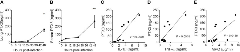
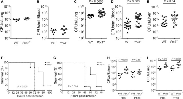
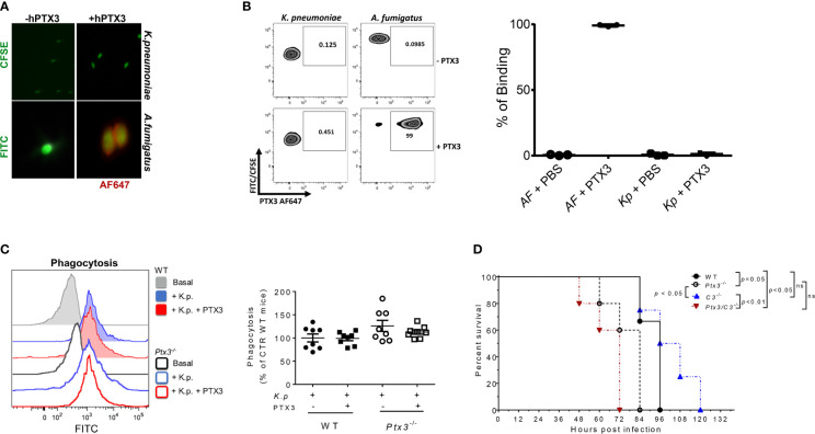
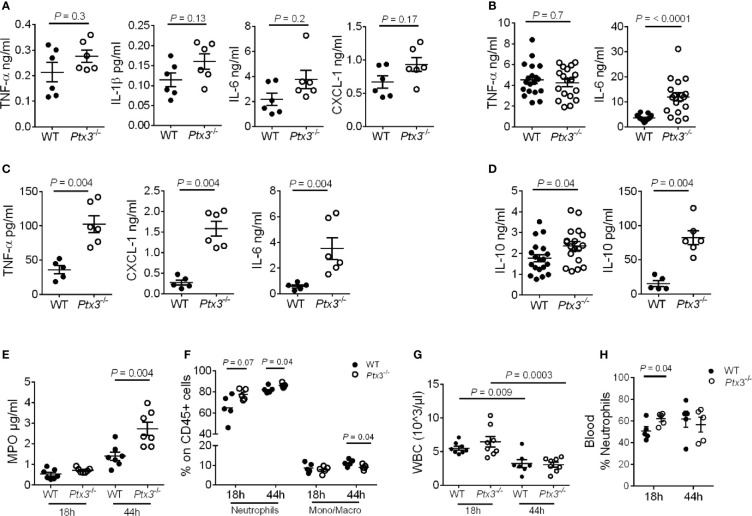
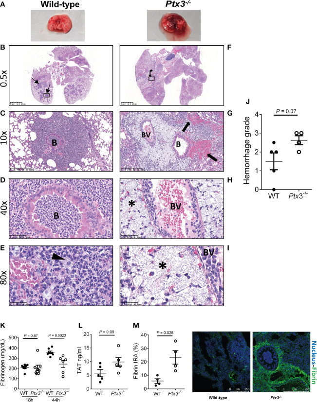
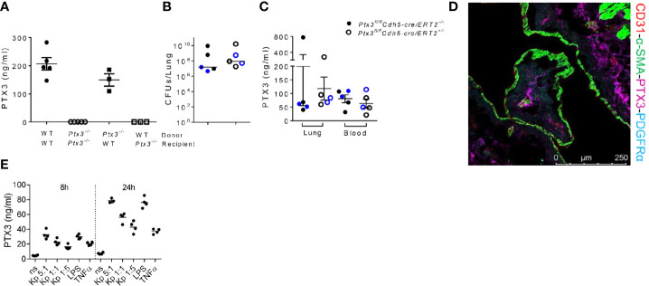
Klebsiella pneumoniae is a common pathogen in human sepsis. The emergence of multidrug-resistant K. pneumoniae strains represents a major clinical challenge in nosocomial and community acquired infections. The long pentraxin PTX3, a key component of humoral innate immunity, is involved in resistance to selected pathogens by promoting opsonophagocytosis. We investigated the relevance of PTX3 in innate immunity against K. pneumoniae infections using Ptx3-/- mice and mouse models of severe K. pneumoniae infections. Local and systemic PTX3 expression was induced following K. pneumoniae pulmonary infection, in association with the up-regulation of TNF-α and IL-1β. PTX3 deficiency in mice was associated with higher bacterial burden and mortality, release of pro-inflammatory cytokines as well as IL-10 in the lung and systemically. The analysis of the mechanisms responsible of PTX3-dependent control of K. pneumoniae infection revealed that PTX3 did not interact with K. pneumoniae, or promote opsonophagocytosis. The comparison of susceptibility of wild-type, Ptx3-/-, C3-/- and Ptx3-/- /C3-/- mice to the infection showed that PTX3 acted in a complement-independent manner. Lung histopathological analysis showed more severe lesions in Ptx3-/- mice with fibrinosuppurative, necrotizing and haemorrhagic bronchopneumonia, associated with increased fibrin deposition in the lung and circulating fibrinogen consumption. These findings indicate that PTX3 contributes to the control of K. pneumoniae infection by modulating inflammatory responses and tissue damage. Thus, this study emphasizes the relevance of the role of PTX3 as regulator of inflammation and orchestrator of tissue repair in innate responses to infections.
Keywords: Klebsiella pneumoniae; Pentraxin 3 (PTX3); complement – immunological term; inflammation; innate immunity; sepsis.
Copyright © 2021 Asgari, Supino, Parente, Polentarutti, Stravalaci, Porte, Pasqualini, Barbagallo, Perucchini, Recordati, Magrini, Mariancini, Riva, Giordano, Davoudian, Roger, Veer, Jaillon, Mantovani, Doni and Garlanda.
Conflict of interest statement
AIM and CG obtain royalties on pentraxin-3 related reagents. The remaining authors declare that the research was conducted in the absence of any commercial or financial relationships that could be construed as a potential conflict of interest.
Figures
Similar articles
-
Acute lung inflammation in Klebsiella pneumoniae B5055-induced pneumonia and sepsis in BALB/c mice: a comparative study.Inflammation. 2011 Oct;34(5):452-62. doi: 10.1007/s10753-010-9253-9. Inflammation. 2011. PMID: 20890649
-
Dual function of the long pentraxin PTX3 in resistance against pulmonary infection with Klebsiella pneumoniae in transgenic mice.Microbes Infect. 2006 Apr;8(5):1321-9. doi: 10.1016/j.micinf.2005.12.017. Epub 2006 Mar 24. Microbes Infect. 2006. PMID: 16697676
-
Platelet and endothelial cell P-selectin are required for host defense against Klebsiella pneumoniae-induced pneumosepsis.J Thromb Haemost. 2015 Jun;13(6):1128-38. doi: 10.1111/jth.12893. Epub 2015 Apr 23. J Thromb Haemost. 2015. PMID: 25773400
-
The Long Pentraxin PTX3 as a Humoral Innate Immunity Functional Player and Biomarker of Infections and Sepsis.Front Immunol. 2019 Apr 12;10:794. doi: 10.3389/fimmu.2019.00794. eCollection 2019. Front Immunol. 2019. PMID: 31031772 Free PMC article. Review.
-
PTX3 Regulation of Inflammation, Hemostatic Response, Tissue Repair, and Resolution of Fibrosis Favors a Role in Limiting Idiopathic Pulmonary Fibrosis.Front Immunol. 2021 Jun 21;12:676702. doi: 10.3389/fimmu.2021.676702. eCollection 2021. Front Immunol. 2021. PMID: 34276664 Free PMC article. Review.
Cited by
-
Novel evidence on sepsis-inducing pathogens: from laboratory to bedside.Front Microbiol. 2023 Jun 23;14:1198200. doi: 10.3389/fmicb.2023.1198200. eCollection 2023. Front Microbiol. 2023. PMID: 37426029 Free PMC article. Review.
-
The role of pentraxin3 in plasma and bronchoalveolar lavage fluid in COPD patients with invasive pulmonary aspergillosis.BMC Pulm Med. 2021 Dec 16;21(1):414. doi: 10.1186/s12890-021-01793-z. BMC Pulm Med. 2021. PMID: 34915889 Free PMC article.
-
Acute phase proteins patterns as biomarkers in bacterial infection: Recent insights.Open Vet J. 2024 Oct;14(10):2539-2550. doi: 10.5455/OVJ.2024.v14.i10.4. Epub 2024 Oct 31. Open Vet J. 2024. PMID: 39545194 Free PMC article. Review.
-
Editorial: Interactions of Pentraxins and Complement in Infection, Inflammation, and Cancer.Front Immunol. 2022 Feb 17;13:861359. doi: 10.3389/fimmu.2022.861359. eCollection 2022. Front Immunol. 2022. PMID: 35251053 Free PMC article. No abstract available.
-
Lactate promotes invasive Klebsiella pneumoniae liver abscess syndrome by increasing capsular polysaccharide biosynthesis via the PTS-CRP axis.Nat Commun. 2025 Jul 1;16(1):6057. doi: 10.1038/s41467-025-61379-9. Nat Commun. 2025. PMID: 40593854 Free PMC article.
References
Publication types
MeSH terms
Substances
LinkOut - more resources
Full Text Sources
Molecular Biology Databases
Miscellaneous