

Visualizing the In Vivo Dynamics of Anti- Leishmania Immunity: Discoveries and Challenges
- PMID: 34093571
- PMCID: PMC8172142
- DOI: 10.3389/fimmu.2021.671582
Visualizing the In Vivo Dynamics of Anti- Leishmania Immunity: Discoveries and Challenges
Abstract
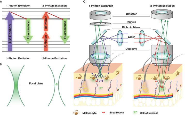
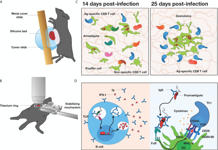
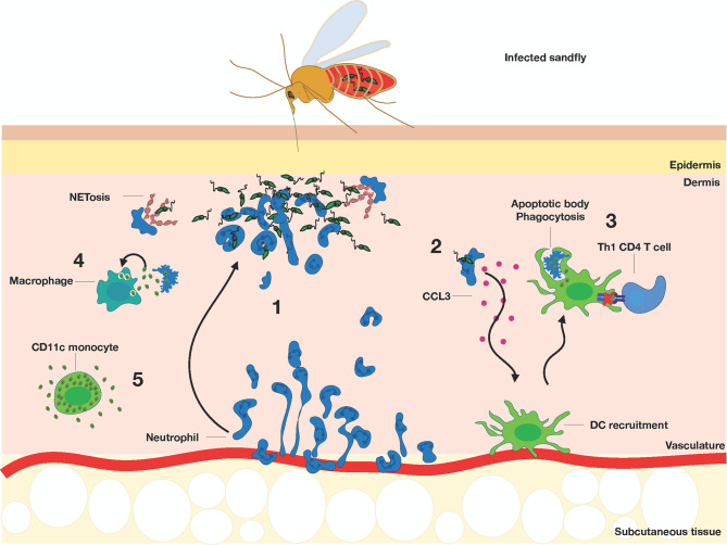
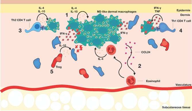
Intravital microscopy, such as 2-photon microscopy, is now a mainstay in immunological research to visually characterize immune cell dynamics during homeostasis and pathogen infections. This approach has been especially beneficial in describing the complex process of host immune responses to parasitic infections in vivo, such as Leishmania. Human-parasite co-evolution has endowed parasites with multiple strategies to subvert host immunity in order to establish chronic infections and ensure human-to-human transmission. While much focus has been placed on viral and bacterial infections, intravital microscopy studies during parasitic infections have been comparatively sparse. In this review, we will discuss how in vivo microscopy has provided important insights into the generation of innate and adaptive immunity in various organs during parasitic infections, with a primary focus on Leishmania. We highlight how microscopy-based approaches may be key to providing mechanistic insights into Leishmania persistence in vivo and to devise strategies for better parasite control.
Keywords: Leishmania infection; T cells; ear skin imaging; fluorescent reporters; liver imaging; macrophages; two-photon intravital microscopy.
Copyright © 2021 Zayats, Uzonna and Murooka.
Conflict of interest statement
The authors declare that the research was conducted in the absence of any commercial or financial relationships that could be construed as a potential conflict of interest.
Figures
References
Publication types
MeSH terms
Grants and funding
LinkOut - more resources
Full Text Sources
Medical