. 2021 May 19:12:672737.
doi: 10.3389/fimmu.2021.672737. eCollection 2021.

Kinetics of Abacavir-Induced Remodelling of the Major Histocompatibility Complex Class I Peptide Repertoire

Affiliations

Kinetics of Abacavir-Induced Remodelling of the Major Histocompatibility Complex Class I Peptide Repertoire

Patricia T Illing et al. Front Immunol. .

Abstract

Abacavir hypersensitivity syndrome can occur in individuals expressing the HLA-B*57:01 major histocompatibility complex class I allotype when utilising the drug abacavir as a part of their anti-retroviral regimen. The drug is known to bind within the HLA-B*57:01 antigen binding cleft, leading to the selection of novel self-peptide ligands, thus provoking life-threatening immune responses. However, the sub-cellular location of abacavir binding and the mechanics of altered peptide selection are not well understood. Here, we probed the impact of abacavir on the assembly of HLA-B*57:01 peptide complexes. We show that whilst abacavir had minimal impact on the maturation or average stability of HLA-B*57:01 molecules, abacavir was able to differentially enhance the formation, selectively decrease the dissociation, and alter tapasin loading dependency of certain HLA-B*57:01-peptide complexes. Our data reveals a spectrum of abacavir mediated effects on the immunopeptidome which reconciles the heterogeneous functional T cell data reported in the literature.

Keywords: MHC I antigen presentation; T cells; abacavir; drug hypersensitivity; immunopeptidome; peptide selection; tapasin.


Conflict of interest statement

The authors declare that the research was conducted in the absence of any commercial or financial relationships that could be construed as a potential conflict of interest.

Figures

Figure 1
Figure 1
Abacavir perturbs the HLA-B*57:01 immunopeptidome by increasing or decreasing the contribution of distinct subsets of peptides. Analysis of 58 selected HLA-B*57:01 peptide ligands, isolated from 108 C1R.B*57:01 cells after 0–16 h, or constant, abacavir treatment using LC-MRM–MS demonstrated four main patterns of impact on contribution to the immunopeptidome; (A) inhibition, (B) minimal impact, (C) facilitated (presented by HLA-B*57:01 of untreated cells but increased in abundance during abacavir treatment) and (D) dependent (only presented by HLA-B*57:01 of cells exposed to abacavir). Perturbation of HLA-B*57:01 ligands increased with time, coincident with increased co-purification of abacavir. Perturbations were not mirrored by the HLA-C ligands analysed (E). Peptide and abacavir abundances are shown as a proportion of the maximum normalised peak area detected and are portrayed as the mean of the three biological replicates (mean +/− SD). The decreased recovery of abacavir at the 6 h point is likely to reflect an experimental artefact in the off line fractionation prior to the LC-MS. Importantly there was not a similar decrease in the amount of peptide material eluted at this time point. Peptides that were chosen for in vitro peptide binding and dissociation experiments are in italics and underlined.
Figure 2
Figure 2
Abacavir does not perturb the proteome of C1R.B*57:01. Comparison of protein abundance between untreated and abacavir treated (48 h, 35 µM) C1R.B*57:01 cells (three replicates per treatment) revealed no major perturbation of the cellular proteome. The volcano plot depicts the difference (log2FC) vs -Logp of 2,306 proteins between untreated and treated conditions. A 5% FDR and a slope of 1 were used as the cut-off for significance (thick black line). No proteins were significantly perturbed by abacavir treatment; furthermore identified source proteins for the HLA ligands analysed clustered close to 0 on the difference axis. Source proteins for HLA ligands from the different subsets analysed are shown as indicated in the key. The purple asterisk shows RACK1, the source protein of peptides KTIKLWNTL (abacavir facilitated), and YTDNLVRVW (minimal impact), and the purple square shows MX1, the source of peptides LTSELITHI (abacavir dependent) and KVVDVVRNL (abacavir inhibited). All other proteins are shown by grey squares.
Figure 3
Figure 3
Immunogenicity of HLA-B*57:01+ antigen presenting cells increases with abacavir exposure time. C1R.B*57:01 cells were cultured in 35 µM abacavir for 0–30 h prior to fixation in paraformaldehyde. Immunogenicity was then gauged by their ability to stimulate cytokine (IFNγ and/or TNF) production in CD8+ abacavir responsive T cells during a 6 h stimulation assay, detected via intracellular cytokine staining. Responses from T cell lines derived from three different HLA-B*57:01+ donors are shown. Donor 1 T cells were assayed in triplicate, whilst donor 2 and 3 T cells were assayed in duplicate in a separate experiment (mean +/− SD).
Figure 4
Figure 4
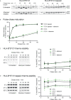
Abacavir does not significantly alter maturation or global stability of HLA-B*57:01. (A) and (B) Maturation [acquisition of resistance to digestion with endoglycosidase-H (Endo-H)] of HLA-B*57:01 molecules was determined by pulse chase in 721.220.B*57:01 or 721.220.B*57:01.tapasin cells, immunoprecipitation of MHC I molecules, and digestion with endoglycosidase-H. (A) depicts representative SDS-PAGE gels for untreated and abacavir treated cells; only the HLA-B*57:01 heavy chain bands are shown. The location of Endo-H resistant (EHR) and Endo-H sensitive (EHS) bands is distinguished by their altered migration pattern. (B) The % EHR material was calculated [EHR/(EHR+EHS)] and plotted as the mean (+/− SD) of three or four replicate experiments for abacavir treated and untreated cells respectively excluding time point 15, which was only included in two experiments. Abacavir had no apparent impact on HLA-B*57:01 maturation in either the presence or absence of tapasin. (C) and (D) Pulse-chase thermostability assays were performed in 721.220.B*57:01 (C) and 721.220.B*57:01.tapasin (D) cells: lysates were prepared from pulse radio-labelled cells and incubated at either 4, 37, or 50°C before β 2m associated MHC I molecules were immunoprecipitated using W6/32 antibody and resolved by SDS-PAGE. Representative SDS-PAGE gels for untreated and abacavir treated cells depict the HLA-B*57:01 heavy chain bands recovered at each chase time after thermal denaturation. Graphs show the amount of HLA-B*57:01 molecules recovered after thermal denaturation at 37 and 50°C as a percentage of that recovered at 4°C (mean +/− SD of four or five replicate experiments for abacavir treated and untreated cells respectively).
Figure 5
Figure 5
Abacavir diversifies the HLA-B*57:01 peptide repertoire by enhancing the loading of specific peptides. Conditional ligand loaded HLA-B*57:01fos molecules were UV exposed before the binding of the indicated peptide (A = KTFK*DVGNLL, B = TSLK*SRVTI, C = LTTK*LTNTNI, D = NTVELRVK*I, E = ATFK*GIVRAI, F = KVFK*LQTSL, G = VTKK*TYEIW, H = ITTK*AISRW, I = RVDPAK*GLFYF) was followed at 25°C in the presence or absence of excess abacavir and/or monomeric or ERp57 C60A conjugated tapasin-jun. Binding of fluorescent peptide is reported in millipolarisation units (mP). Unbound fluorescent peptide is assumed to have an mP level of 50. Data shown are representative of triplicate or greater experiments. The extent that tapasin, abacavir, or the combination of both enhanced the binding of fluorescent peptides to HLA-B*57:01fos molecules in the replicate experiments is presented in Figure 6 . Colour coding is as follows: grey—peptide alone, blue—abacavir, red—tapasin, green—abacavir + tapasin.
Figure 6
Figure 6
Tapasin and abacavir differentially enhance the binding of fluorescent peptides. The extent that (A) tapasin, (B) abacavir, or (C) the combination of both enhanced the binding of fluorescent peptides to HLA-B*57:01fos molecules was compared over independent experiments. For each polarisation measurement taken in an experiment the enhancement factor was calculated by: dividing polarisation in the presence of tapasin (or abacavir or both) by the intrinsic polarisation level; multiplying by 100 to obtain a percentage; and calculating the mean enhancement for the experiment. The enhancement factors obtained from different experiments are shown as dots, with the mean average shown as a horizontal black bar, and the standard deviation of multiple experiments shown as a coloured vertical line edged with horizontal bars. Peptides are coloured according to their classification by mass spectrometry: abacavir facilitated = yellow, abacavir dependent = green, minimally affected = orange, abacavir inhibited = red.
Figure 7
Figure 7
Abacavir diversifies the HLA-B*57:01 peptide repertoire by decreasing dissociation of specific peptides. Conditional ligand loaded HLA-B*57:01fos molecules were UV exposed and incubated with the indicated fluorescent peptide (A = VTKK*TYEIW, B = RVDPAK*GLFYF, C = KVFK*LQTSL, D = NTVELRVK*I, E = ATFK*GIVRAI, F = KTFK*DVGNLL, G = TSLK*SRVTI, H = ITTK*AISRW). Fluorescence polarisation measurements were taken after the addition of excess unlabelled competing peptide in the absence or presence of abacavir and/or conjugated tapasin-jun-ERp57 C60A proteins. Data shown are representative of triplicate or greater experiments.
Figure 8
Figure 8
Proposed model of peptide-loading of HLA-B*57:01 in the presence of abacavir. (A) When abacavir is present, abacavir-dependent peptides are loaded into HLA-B*57:01 within the endoplasmic reticulum (left). For the abacavir-dependent peptides in our panel, loading was highly dependent on abacavir as compared to tapasin, represented by the scales tipping towards abacavir (magenta, A), as opposed to tapasin (teal, T). After a slight delay consistent with de novo complex generation in the ER and progression through the secretory pathway, this generates conformationally novel HLA–abacavir–peptide complexes at the cell surface (right, novel self-peptide and novel conformation). The inset box represents the increasing appearance of cell surface complexes (0%–max) incorporating abacavir-dependent peptides (green) and abacavir (magenta hatching) over time (blue arrow) after abacavir addition (pink arrow), until a maximum is reached (not to scale). (B) Abacavir-facilitated peptides are part of the constitutive immunopeptidome (upper panel). For the abacavir-facilitated peptides in our panel, tapasin aids loading in the endoplasmic reticulum in the absence of abacavir, and peptide-loaded HLA traffic to the cell surface. On addition of abacavir, abacavir can load into these HLA-peptide complexes at the cell surface generating novel conformations/stabilising the structure (constitutive peptide, novel conformation), contributing immediately to cellular immunogenicity. In addition, de novo generation of HLA-abacavir-peptide complexes occurs within the endoplasmic reticulum, with both abacavir and tapasin promoting peptide binding (lower panel). As such abacavir-facilitated peptides are present in the immunopeptidome prior to abacavir addition (inset box, yellow), with immediate loading of abacavir at the cell surface as well as during de novo complex generation, contributing an increasing number of abacavir occupied HLA from the time of abacavir addition (inset box, magenta hatching). (C) These changes occur against a background of constitutive peptides which maintain a similar contribution to the immunopeptidome regardless of the presence of abacavir (minimal impact, inset box, orange), whilst a proportion of constitutive peptides are reduced in presentation (inhibited, Figure 1 , not shown here). This figure was created with BioRender.com.

Similar articles

Cited by

References

    1. Yewdell JW, Anton LC, Bennink JR. Defective Ribosomal Products (DriPs): A Major Source of Antigenic Peptides for MHC Class I Molecules? J Immunol (1996) 157(5):1823–6. - PubMed
    1. Zernich D, Purcell AW, Macdonald WA, Kjer-Nielsen L, Ely LK, Laham N, et al. . Natural HLA Class I Polymorphism Controls the Pathway of Antigen Presentation and Susceptibility to Viral Evasion. J Exp Med (2004) 200(1):13–24. 10.1084/jem.20031680 - DOI - PMC - PubMed
    1. Williams AP, Peh CA, Purcell AW, McCluskey J, Elliott T. Optimization of the MHC Class I Peptide Cargo is Dependent on Tapasin. Immunity (2002) 16(4):509–20. 10.1016/S1074-7613(02)00304-7 - DOI - PubMed
    1. Turnquist HR, Thomas HJ, Prilliman KR, Lutz CT, Hildebrand WH, Solheim JC. HLA-B Polymorphism Affects Interactions With Multiple Endoplasmic Reticulum Proteins. Eur J Immunol (2000) 30(10):3021–8. 10.1002/1521-4141(200010)30:10<3021::AID-IMMU3021>3.0.CO;2-U - DOI - PubMed
    1. Fang Q, Liu Z, Zhang T. Human Leukocyte Antigen Polymorphisms and Kaposi’s Sarcoma-Associated Herpesvirus Infection Outcomes: A Call for Deeper Exploration. J Med Virol (2019) 91(4):541–8. 10.1002/jmv.25342 - DOI - PubMed

Publication types