. 2021 May 20:12:680568.
doi: 10.3389/fimmu.2021.680568. eCollection 2021.

CD47 Deficiency Ameliorates Ocular Autoimmune Inflammation

Affiliations

CD47 Deficiency Ameliorates Ocular Autoimmune Inflammation

Yoko Okunuki et al. Front Immunol. .

Abstract

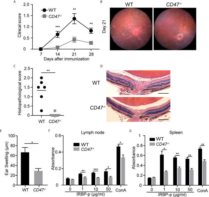
Autoimmune uveitis is a sight-threatening ocular inflammatory condition in which the retina and uveal tissues become a target of autoreactive immune cells. The CD47 is a ubiquitously expressed transmembrane protein which plays multiple roles in fundamental cellular functions including phagocytosis, proliferation, and adhesion. Signal regulatory protein alpha (SIRPα), one of the CD47 ligands, is predominantly expressed in myeloid lineage cells such as dendritic cells (DCs) or macrophages, and CD47-SIRPα signaling pathway is implicated in the development of autoimmune diseases. Our current study demonstrates how CD47 depletion is effective in the prevention of experimental autoimmune uveitis (EAU), an animal model of human autoimmune uveitis, in animals deficient of CD47 (CD47-/- ). Systemic suppression of SIRPα+ DCs in animals deficient in CD47 resulted in the inability of autoreactive CD4+ T cells to develop, which is crucial to induction of EAU. Of interest, retinal microglia, the resident immune cell of the retina, express SIRPα, however these cells were not operative in EAU suppression in response to CD47 depletion. These results identify CD47 as a significant regulator in the development of SIRPα+ DCs that is vital to disease induction in EAU.

Keywords: CD47 antigen; antigen-presenting cells; autoimmune diseases; retina; uveitis.


Conflict of interest statement

The authors declare that the research was conducted in the absence of any commercial or financial relationships that could be construed as a potential conflict of interest.

Figures

Figure 1
Figure 1
CD47 deficient mice are protected from autoimmune uveitis. Female WT and CD47-/- mice were immunized with IRBP-p and the animals were evaluated for (A–D) severity of retinal inflammation and (E–G) systemic immune responses against the IRBP-p. (A) Time course of EAU clinical scores were evaluated by fundus observation (n =10-11 mice per group). (B) Representative retinal fundus images on day 21. (C) Histopathological EAU score was evaluated on day 21 (n = 6 mice per group). (D) Representative histopathological (H&E staining) images (Scale bars: 200 μm). (A, C) Data were analyzed by Mann–Whitney U test. (E) Delayed hypersensitivity, as determined by ear swelling, was evaluated on day 21 (n=10-11). (F, G) Cell proliferation was evaluated by using the cells isolated from lymph nodes and spleens on day 21. The cells were cultured in triplicate for 3 days in the presence of IRBP-p (10 μg/ml), Con A (1 μg/ml), or medium only (n = 5 mice per group). (E–G) Data were analyzed by Student’s t test. Data are expressed as mean ± SEM. *P < 0.05; **P < 0.01; ***P < 0.001.
Figure 2
Figure 2
SIRPα expression on retinal microglia is upregulated in naïve CD47-/- mice. Retinal microglia are isolated from naïve WT and CD47-/- mice. Both retinas from one mouse were combined and processed as one sample. Retinal single cell suspensions were gated on live cells (DAPI-). SIRPα expression on microglia (CD11b+CD45low) was examined by flow cytometry. (A) The gating strategy of microglia and SIRPα, and representative sample from each group. (B) MFI of SIRPα on microglia. (C) The number of microglia per sample (contains two retinas). Data were analyzed by Student’s t test and expressed as mean ± SEM. ****P<0.0001. n.s., not significant. n=4 mice per group.
Figure 3
Figure 3
CD47 deficiency in systemic immune cells but not locally in the eye is protective for autoimmune uveitis. Time course of clinical score and representative fundus images using adoptive transfer EAU model. (A, B) WT donor mice were immunized and the primed cells were transferred to naive WT or CD47-/- recipient mice. (A) Time course of EAU clinical scores were evaluated by fundus observation (n =4 mice per group) (B) Representative retinal fundus images on day 13. (C, D) WT and CD47-/- donor mice were immunized and the primed cells were transferred to naive WT recipient mice. (C) Time course of EAU clinical scores were evaluated by fundus observation (n=5-6 mice per group) (D) Representative retinal fundus images on day 17. Data were analyzed by MannWhitney U test and expressed as mean ± SEM. **P<0.01; n.s., not significant.
Figure 4
Figure 4
The number of T cells, macrophages, and DCs are decreased in CD47 deficient mice with autoimmune uveitis. Spleen and lymph node cells from WT and CD47-/- mice were collected before (naive), and 14 days and 21 days post immunization. (A) Total cell number in spleens and lymph nodes. (B) CD4+, CD8+, CD19+, CD11b+, CD11c+ cells in spleens and lymph nodes were detected by flowcytometry and cell number of each population was calculated. The cells were gated on live CD45+CD3+ cells to detect CD4+ T cells and CD8+ T cells. CD45+CD3- population was further gated on CD19, CD11b, and CD11c. Data were analyzed by Student’s t test and expressed as mean ± SEM. *P<0.05; **P<0.01; ***P<0.001. n.s., not significant. n=4 mice per group.
Figure 5
Figure 5
SIRPα expression in CD11b+ macrophages/DCs is decreased in CD47 deficient mice with autoimmune uveitis. WT and CD47-/- mice were immunized and spleen cells were collected 14 days post immunization and examined by flow cytometry. (A) Live cells (Zombie UV-) excluding a dump gate (T cells; CD3, B cells; CD19, NK cells; NK1.1, and granulocytes; Ly6G) were gated on CD45+ cells and further separated by CD11b and CD11c. The gates were defined as follows: Gates a and a’; CD11b+CD11chigh (CD11b+ DCs), gates b and b’; CD11b+CD11c- (macrophages), gates c and c’; CD11blowCD11c+ (CD11blow DCs), and gates d and d’; CD11b-CD11clow (CD11b- DCs). (B) SIRPαhigh cells and CD8a+ cells in (A) are shown in green and blue, respectively. (C) SIRPα expression on each macrophage and DC population gated in (A) was examined. (D) Frequency of each macrophage/DC population in spleens was determined by using frequency of each population in ZombieUV-dump-CD45+ cells. (E) Cell number of each macrophage/DC population was obtained by using the number of cells in each SP and the frequency of each population in ZombieUV- cells. Data were analyzed by Student’s t test and expressed as mean ± SEM. *P<0.05; **P<0.01; ***P<0.001. n.s., not significant. n=4 mice per group.
Figure 6
Figure 6
Activation of macrophages and DCs is suppressed in EAU in CD47 deficient mice. WT and CD47-/- mice were immunized and SP and LN cells were collected 14 days post immunization. Expression of cell activation makers on CD4+ T cells, and CD11b+ (DCs/macrophages) cells and CD11c+ (DCs) cells were evaluated by flow cytometry. (A, B) For CD4+ T cells, live cells excluding B cell (CD19), NK cells (NK1.1), and granulocytes (Ly6G) were gated on CD45+CD3+CD4+ cells. Frequency of CD62-CD44+ (effector) cells, CD62+CD44- (naïve) cells, and CD25+ activated CD4+ T cells were examined in spleens (A) and lymph nodes (B). (C, D) For DCs/macrophages, live cells excluding T cells (CD3), B cells (CD19), NK cells (NK1.1), and granulocytes (Ly6G) were gated on CD45+CD11b+ and CD45+CD11c+ cells. Expression of activation markers CD80, CD86, and MHC-II was examined in spleens (C) and lymph nodes (D). Data were analyzed by Student’s t test and expressed as mean ± SEM. *P<0.05; **P<0.01; ***P<0.001. n.s., not significant. n=4 mice per group.
Figure 7
Figure 7
DCs from CD47 deficient mice failed to stimulate T cells in antigen specific response. WT and CD47-/- mice (n=3-4) were immunized and SP and LN cells were collected 14 days post immunization. CD4+ T cells and DCs (CD11c+) purified from pooled LN cells and SP cells, respectively, were incubated at a ratio of T cells: DCs =2:1 (2 × 10⁵: 1 × 10⁵/well) in the presence of 10 μg/ml IRBP-p in a 96 well U-bottom plate for 3 days. (A) Proliferated cells with reduced expression of CellTrace™ Far Red in live CD3+ CD4+ cells were analyzed by flowcytometry. (B) Representative flow cytometry plots. The samples were run triplicates. Data were analyzed by one-way ANOVA followed by Tukey’s multiple comparison. Data are expressed as mean ± SEM. **P<0.01; ***P<0.001.

Similar articles

Cited by

References

    1. Goto H, Mochizuki M, Yamaki K, Kotake S, Usui M, Ohno S. Epidemiological Survey of Intraocular Inflammation in Japan. Jpn J Ophthalmol (2007) 51:41–4. 10.1007/s10384-006-0383-4 - DOI - PubMed
    1. Durrani OM, Tehrani NN, Marr JE, Moradi P, Stavrou P, Murray PI. Degree, Duration, and Causes of Visual Loss in Uveitis. Br J Ophthalmol (2004) 88:1159–62. 10.1136/bjo.2003.037226 - DOI - PMC - PubMed
    1. Rosenbaum JT, Bodaghi B, Couto C, Zierhut M, Acharya N, Pavesio C, et al. . New Observations and Emerging Ideas in Diagnosis and Management of non-Infectious Uveitis: A Review. Semin Arthritis Rheum (2019) 49:438–45. 10.1016/j.semarthrit.2019.06.004 - DOI - PubMed
    1. Kunimi K, Usui Y, Tsubota K, Mitsuhashi R, Umazume A, Kezuka T, et al. . Changes in Etiology of Uveitis in a Single Center in Japan. Ocul Immunol Inflamm (2020) 1–6. 10.1080/09273948.2019.1709649 - DOI - PubMed
    1. Lee RW, Nicholson LB, Sen HN, Chan CC, Wei L, Nussenblatt RB, et al. . Autoimmune and Autoinflammatory Mechanisms in Uveitis. Semin Immunopathol (2014) 36:581–94. 10.1007/s00281-014-0433-9 - DOI - PMC - PubMed

Publication types

MeSH terms