. 2021 May 20:2021:6663977.
doi: 10.1155/2021/6663977. eCollection 2021.

Pixel-Wise Classification in Hippocampus Histological Images

Affiliations

Pixel-Wise Classification in Hippocampus Histological Images

Alfonso Vizcaíno et al. Comput Math Methods Med. .

Abstract

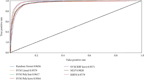
This paper presents a method for pixel-wise classification applied for the first time on hippocampus histological images. The goal is achieved by representing pixels in a 14-D vector, composed of grey-level information and moment invariants. Then, several popular machine learning models are used to categorize them, and multiple metrics are computed to evaluate the performance of the different models. The multilayer perceptron, random forest, support vector machine, and radial basis function networks were compared, achieving the multilayer perceptron model the highest result on accuracy metric, AUC, and F 1 score with highly satisfactory results for substituting a manual classification task, due to an expert opinion in the hippocampus histological images.


Conflict of interest statement

The authors declare that they have no conflicts of interest.

Figures

Figure 1
Figure 1
Coronal sections of the rat brain where the layers of the hippocampus stained with hematoxylin and eosin are observed. 3x magnification. Columns show difference in colour as well as in lighting. Rows show variability in hippocampus shape and markings presence.
Figure 2
Figure 2
Diagram of the implemented methodology for hippocampus pixel-wise classification.
Figure 3
Figure 3
(a) Original image. (b) Final image: rotated, centred, and resized.
Figure 4
Figure 4
(a, d) Colour independence. (b, e) Background homogenization. (c, f) Hippocampus enhancement.
Figure 5
Figure 5
A hippocampus section is framed with cyan colour. Other brain regions that have a similar shape and similar pixel characteristics are framed with green colour.
Figure 6
Figure 6
Red dots represent hippocampus pixels and blue dots represent nonhippocampus pixels. (a) Sample image from training set, (b) the same sample image taken from validation set, and (c) the same sample image taken from test set.
Figure 7
Figure 7
ROC curve. Dotted line from bottom left to upper right represents a strictly random classifier.

References

    1. Norman K. A. How hippocampus and cortex contribute to recognition memory: revisiting the complementary learning systems model. Hippocampus. 2010;20(11):1217–1227. doi: 10.1002/hipo.20855. - DOI - PMC - PubMed
    1. Golsorkhdan S. A., Boroujeni M. E., Aliaghaei A., et al. Methamphetamine administration impairs behavior, memory and underlying signaling pathways in the hippocampus. Behavioural Brain Research. 2020;379, article 112300 doi: 10.1016/j.bbr.2019.112300. - DOI - PubMed
    1. Barnes J., Bartlett J. W., van de Pol L. A., et al. A meta-analysis of hippocampal atrophy rates in Alzheimer's disease. Neurobiology of Aging. 2009;30(11):1711–1723. doi: 10.1016/j.neurobiolaging.2008.01.010. - DOI - PMC - PubMed
    1. Kaya S., Yildirim H., Atmaca M. Reduced hippocampus and amygdala volumes in antisocial personality disorder. Journal of Clinical Neuroscience. 2020;75:199–203. doi: 10.1016/j.jocn.2020.01.048. - DOI - PubMed
    1. Bouët V., Freret T., Toutain J., Divoux D., Boulouard M., Schumann-Bard P. Sensorimotor and cognitive deficits after transient middle cerebral artery occlusion in the mouse. Experimental Neurology. 2007;203(2):555–567. doi: 10.1016/j.expneurol.2006.09.006. - DOI - PubMed

MeSH terms

LinkOut - more resources