

Clinical value of color Doppler ultrasound combined with serum tumor markers for the diagnosis of medullary thyroid carcinoma
- PMID: 34093776
- PMCID: PMC8170263
- DOI: 10.3892/ol.2021.12822
Clinical value of color Doppler ultrasound combined with serum tumor markers for the diagnosis of medullary thyroid carcinoma
Abstract
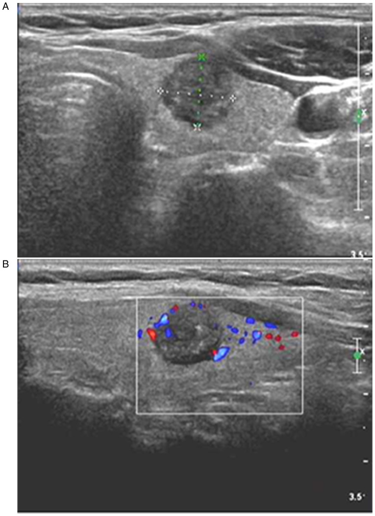
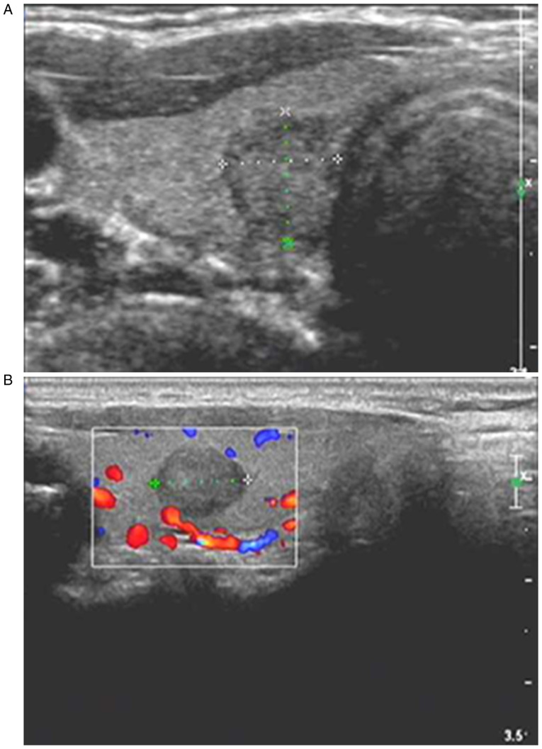
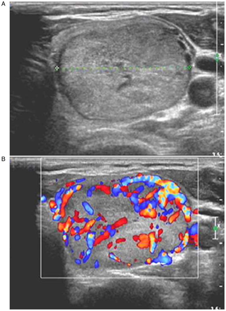
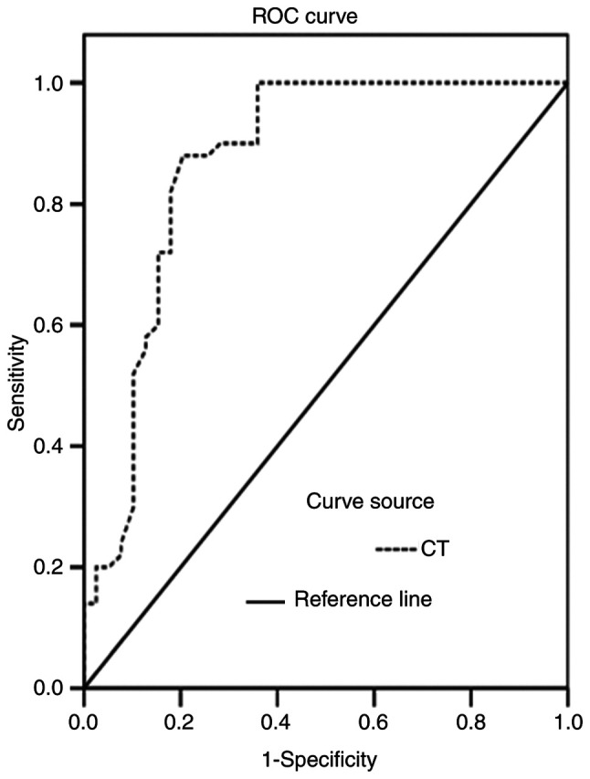
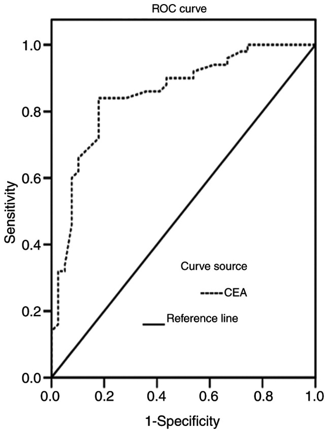
The present study aimed to explore the clinical value of color Doppler ultrasound combined with serum tumor markers, including calcitonin (CT) and carcinoembryonic antigen (CEA), for the diagnosis of medullary thyroid carcinoma (MTC). A total of 39 patients with MTC (MTC group), 50 patients with papillary thyroid carcinoma (PTC) (PTC group) and 30 patients with thyroid adenoma (benign control group) were enrolled in the present study. The patients were hospitalized at the Affiliated Hospital of Qingdao University from January 2012 to December 2018 and were diagnosed through surgical procedures and pathology laboratory results. The ultrasound results, as well as serum CT and CEA results, were collected and analyzed. A significant difference was observed between the MTC and PTC groups in regards to morphology, margin, aspect ratio, calcification, internal blood flow and lymph node metastasis (all P<0.01). There was also a significant difference between the MTC and benign control group in regards to internal echo, calcification, internal blood flow and lymph node metastasis (all P<0.01). In addition, the levels of serum CT and CEA in the MTC group were significantly higher than those in the PTC and the benign control groups (both P<0.01). For patients with MTC, the levels of serum CT and CEA were significantly associated with maximum tumor diameter, lymph node metastasis and the patient state after treatment (all P<0.01). Furthermore, the sensitivities of ultrasound, serum CT and CEA for the diagnosis of MTC were 76.92, 74.36 and 68.23%, respectively. The value for the combination of the three markers (94.87%) was significantly higher compared with the sensitivity value of each separate marker (all P<0.05). In conclusion, color Doppler ultrasound combined with detecting the levels of serum tumor markers (CT and CEA) significantly improved the diagnostic efficiency for MTC, which could be useful for the clinical diagnosis and treatment of MTC.
Keywords: carcinoembryonic antigen; color Doppler ultrasound; joint examination; medullary thyroid carcinoma; serum calcitonin.
Copyright: © Yang et al.
Conflict of interest statement
The authors declare that they have no competing interests.
Figures
Similar articles
-
Diagnostic value of preoperative systemic inflammatory markers and carcinoembryonic antigen in medullary thyroid carcinoma and the risk factors affecting its prognosis.Gland Surg. 2025 Jan 24;14(1):13-27. doi: 10.21037/gs-24-397. Epub 2025 Jan 20. Gland Surg. 2025. PMID: 39958900 Free PMC article.
-
[Ultrasonographic features of thyroid carcinoma of different sizes: comparison between medullary thyroid carcinomas and papillary thyroid carcinomas].Zhonghua Zhong Liu Za Zhi. 2024 Feb 23;46(2):133-139. doi: 10.3760/cma.j.cn112152-20231026-00264. Zhonghua Zhong Liu Za Zhi. 2024. PMID: 38418187 Chinese.
-
Measuring discrepancies between simple medullary and synchronous medullary/papillary thyroid carcinomas: a comparative cross-sectional study.Front Endocrinol (Lausanne). 2024 Jan 22;14:1301200. doi: 10.3389/fendo.2023.1301200. eCollection 2023. Front Endocrinol (Lausanne). 2024. PMID: 38317715 Free PMC article. Review.
-
Combining serum calcitonin, carcinoembryonic antigen, and neuron-specific enolase to predict lateral lymph node metastasis in medullary thyroid carcinoma.J Clin Lab Anal. 2020 Jul;34(7):e23278. doi: 10.1002/jcla.23278. Epub 2020 Mar 6. J Clin Lab Anal. 2020. PMID: 32141647 Free PMC article.
-
A Review of the Significance in Measuring Preoperative and Postoperative Carcinoembryonic Antigen (CEA) Values in Patients with Medullary Thyroid Carcinoma (MTC).Medicina (Kaunas). 2021 Jun 11;57(6):609. doi: 10.3390/medicina57060609. Medicina (Kaunas). 2021. PMID: 34208296 Free PMC article. Review.
Cited by
-
Preoperative Serum Calcitonin Level and Ultrasonographic Characteristics Predict the Risk of Metastatic Medullary Thyroid Carcinoma: Functional Analysis of Calcitonin-Related Genes.Dis Markers. 2022 Mar 2;2022:9980185. doi: 10.1155/2022/9980185. eCollection 2022. Dis Markers. 2022. PMID: 35280443 Free PMC article.
-
Inorganic Nanomaterial for Biomedical Imaging of Brain Diseases.Molecules. 2021 Dec 3;26(23):7340. doi: 10.3390/molecules26237340. Molecules. 2021. PMID: 34885919 Free PMC article. Review.
-
Holomics and Artificial Intelligence-Driven Precision Oncology for Medullary Thyroid Carcinoma: Addressing Challenges of a Rare and Aggressive Disease.Cancers (Basel). 2024 Oct 13;16(20):3469. doi: 10.3390/cancers16203469. Cancers (Basel). 2024. PMID: 39456563 Free PMC article. Review.
-
Radiomics Features of Different Sizes of Medullary Thyroid Carcinoma (MTC) and Papillary Thyroid Carcinoma (PTC) Tumors: A Comparative Study.Clin Med Insights Oncol. 2022 May 15;16:11795549221097675. doi: 10.1177/11795549221097675. eCollection 2022. Clin Med Insights Oncol. 2022. PMID: 35603093 Free PMC article.
-
Can ACR TI-RADS predict the malignant risk of medullary thyroid cancer?J Clin Transl Endocrinol. 2024 Dec 20;39:100380. doi: 10.1016/j.jcte.2024.100380. eCollection 2025 Mar. J Clin Transl Endocrinol. 2024. PMID: 39811784 Free PMC article.
References
LinkOut - more resources
Full Text Sources