. 2021 May 5;12(13):3900-3908.
doi: 10.7150/jca.54574. eCollection 2021.

Wighteone exhibits an antitumor effect against EGFR L858R/T790M mutation non-small cell lung cancer

Affiliations

Wighteone exhibits an antitumor effect against EGFR L858R/T790M mutation non-small cell lung cancer

Peiyuan Sun et al. J Cancer. .

Abstract

Non-small cell lung cancer (NSCLC) harboring activating EGFR mutations were initially treated by first-generation EGFR tyrosine kinase inhibitors (EGFR-TKIs), unfortunately, the efficacy of these drugs is limited, mostly frequent due to T790M mutation. Although osimertinib has been approved to treat patients with T790M-positive NSCLC, the majority of patients will develop C797S mutation and suffer diseases again. Therefore, more novel therapeutic strategies for T790M mutation-positive NSCLC are urgently required. We hypothesized that wighteone, a natural compound isolated from plant derivatives, has antitumor effects against NSCLC with T790M mutation. In this study, we created a Ba/F3 cell line harboring EGFR L858R/T790M mutation (Ba/F3 EGFR L858R/T790M cell line), and then used this cell line and a human NSCLC cell line with EGFR L858R/T790M mutation (NCI-H1975) to investigate the effects and mechanism of wighteone. The results showed that wighteone inhibited cell proliferation, suppressed EGFR signaling pathway, caused cell cycle redistribution and induced cell apoptosis. Our studies suggest that wighteone may provide a novel potential therapeutic strategy for NSCLC patients with T790M mutation.

Keywords: EGFR; L858R/T790M mutation; NSCLC; antitumor effect; wighteone.


Conflict of interest statement

Competing Interests: The authors have declared that no competing interest exists.

Figures

Figure 1
Figure 1
A stable Ba/F3 EGFR L858R/T790M cell line was constructed. (A) After electroporation and monoclone, the EGFR expression levels of transfected cells were detected by flow cytometry. Unstained cells were denoted by black region, while cells stained with specific anti-EGFR antibody were shown as green region. (B) IL-3-independent growth assay of Ba/F3 and Ba/F3 EGFR L858R/T790M cells was evaluated. Values for cells grown without IL-3 were normalized to the values for cells grown with IL-3. (C) After cells were treated with various concentration of EGF, the expression levels of p-EGFR and EGFR were determined by Western blot. Data represent the average of three independent experiments (mean ± SEM). *** p < 0.001 vs + IL-3.
Figure 2
Figure 2
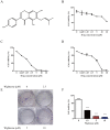
Wighteone potently inhibited EGFRL858R/T790M. (A) Chemical structure of wighteone. Inhibitory effects of wighteone were evaluated in Ba/F3 (B), Ba/F3 EGFR L858R/T790M (C) and NCI-H1975 (D) cells. (E) NCI-H1975 cells were treated with different concentrations of wighteone and colony efficiency was observed by a colony formation assay. (F) Quantitative results of clonogenic effects were analyzed. Data represent the average of three independent experiments (mean ± SEM). *** p < 0.001 vs the control.
Figure 3
Figure 3
Wighteone suppressed the EGFR signaling pathway in NCI-H1975 cells. (A) NCI-H1975 cells were stimulated with EGF (20ng/ml) for 5 minutes and further treated with various concentrations of wighteone (2.5, 5, or 10 µM) for 16h. The expression levels of relative proteins were detected by Western blot with β-tubulin as the loading control. (B-D) The p-EGFR, p-Erk and p-AKT levels were quantified as percentage versus their relative total protein levels. Data represent the average of three independent experiments (mean ± SEM). ### p < 0.001 vs the control; ** p < 0.01, *** p < 0.001 vs EGF.
Figure 4
Figure 4
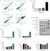
Wighteone regulates cell cycle in NCI-H1975 cells. (A) The cell cycle distribution (sub-G1, G0/G1, S, and G2/M) was detected by flow cytometry. NCI-H1975 cells were treated with various concentrations of wighteone for 24h. (B) The proportions of cells in each phase were quantified as percentages. (C) After NCI-H1975 cells were treated with EGF for 5 min and then further treated with wighteone for 16 h, the expression levels of CDK2, cyclin A and cyclin E were determined by Western blot, using GAPDH as the loading control. (D-F) CDK2, cyclin A and cyclin E protein levels were quantified. Data represent the average of three independent experiments (mean ± SEM). ### p < 0.001 vs the control; * p < 0.05, *** p < 0.001 vs EGF.
Figure 5
Figure 5
Wighteone induces cell apoptosis in NCI-H1975 cells. (A) Flow cytometry was used to detect cell apoptosis in NCI-H1975 cells treated with various concentrations of wighteone. (B) Quantitative results of apoptotic cells in each group were quantified as percentages. (C) The expression levels of relative protein were determined by Western blot, using β-tubulin as the loading control. Then, cleaved-caspase9 (D), cleaved-caspase3 (E), cleaved-PARP1 (F), Bcl-2 (G) and Bax (H) protein levels were analyzed. Data represent the average of three independent experiments (mean ± SEM). ## p < 0.01, ### p < 0.001 vs the control; * p < 0.05, ** p < 0.01, *** p < 0.001 vs EGF.
Figure 6
Figure 6
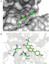
In docking model of EGFR L858R/T790M mutant (PDB code: 3W2P) interaction with wighteone. (A) Binding of wighteone to the ATP-binding domain of EGFRL858R/T790M. EGFRL858R/T790M was depicted by a hydrophobicity surface model (grey). (B) Molecular docking model of wighteone bound to EGFRL858R/T790M. Ligand and key residues are shown as sticks (C, yellow; O, red; N, blue; S, yellow; polar H, white), while hydrogen bonds are denoted by yellow dashed lines.

Similar articles

Cited by

References

    1. Travis WD, Brambilla E, Nicholson AG. et al. The 2015 World Health Organization classification of lung tumors: impact of genetic, clinical and radiologic advances since the 2004 classification. J Thorac Oncol. 2015;10:1243–60. - PubMed
    1. Siegel RL, Miller KD, Jemal A. Cancer statistics, 2019. CA Cancer J Clin. 2019;69:7–34. - PubMed
    1. Chen Z, Fillmore CM, Hammerman PS. et al. Non-small cell lung cancers: a heterogeneous set of diseases. Nat Rev Cancer. 2014;14:535. - PMC - PubMed
    1. Torre LA, Bray F, Siegel RL. et al. Global cancer statistics, 2012. CA Cancer J Clin. 2015;65:87–108. - PubMed
    1. Yoshida T, Zhang G, Haura EB. Targeting epidermal growth factor receptor: central signaling kinase in lung cancer. Biochem Pharmacol. 2010;80:613–23. - PubMed