

MicroRNA-1246 by Targeting AXIN2 and GSK-3β Overcomes Drug Resistance and Induces Apoptosis in Chemo-resistant Leukemia Cells
- PMID: 34093820
- PMCID: PMC8176421
- DOI: 10.7150/jca.58522
MicroRNA-1246 by Targeting AXIN2 and GSK-3β Overcomes Drug Resistance and Induces Apoptosis in Chemo-resistant Leukemia Cells
Abstract
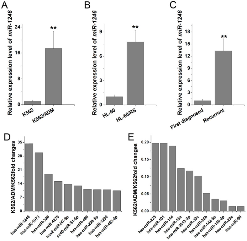
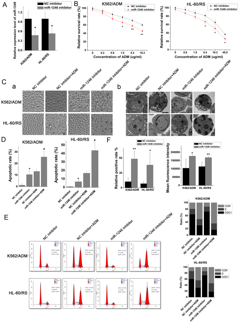
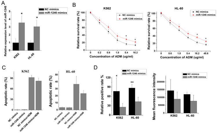
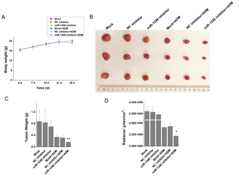
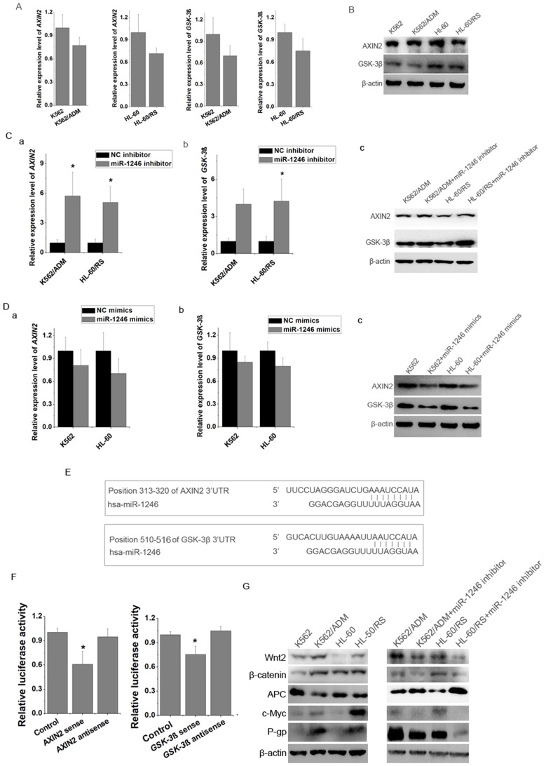
Background and objective: Chemotherapy plays an important role in the treatment of leukemia. Multidrug resistance (MDR) induced by chemotherapy always leads to treatment failure and disease recurrence. MicroRNAs (miRNAs) have been verified as crucial components in carcinogenesis, including chemo-resistance of tumor cells, which has not been fully understood. In this study, we aimed to identify the potential candidate miRNA, miR-1246, and reveal its regulatory role in chemo-resistance of leukemia cells. Methods: Candidate miRNAs were selected by microarray analysis, screened by bioinformatics tools and verified by reverse transcription-quantitative polymerase chain reaction (RT-qPCR). Chemo-resistant phenotypes, including cell viability, apoptosis, adriamycin (ADM) efflux and in vivo oncogenicity of leukemia cells following transfected with miR-1246 mimics or inhibitor were checked with or without ADM treatment to make clear the relationship between miR-1246 and chemo-resistance. RT-qPCR, western blot and dual luciferase reporter assay were performed to measure the expression of related genes and address the potential regulatory mechanism of miR-1246 in chemo-resistance. Results: The expression of miR-1246 was significantly higher in chemo-resistant leukemia K562/ADM cells, HL-60/RS cells and recurrent primary leukemia cells. Loss of miR-1246 inhibited proliferation, induced apoptosis, altered cell cycle distribution, inhibited ADM efflux in chemo-resistant leukemia cells, while overexpression of miR-1246 showed the opposite role in chemo-sensitive leukemia cells. Both bioinformatics prediction and luciferase assay indicated that AXIN2 and glycogen synthase kinase 3 beta (GSK-3β) were the direct targets of miR-1246 in leukemia cells. Inhibition of miR-1246 could up-regulate AXIN2 and GSK-3β and inactivate Wnt/β-catenin pathway, accompanied with inhibiting the expression of β-catenin and further influencing the expression of P-glycoprotein (P-gp) in the chemo-resistant leukemia cells. Conclusions: Chemo-resistant ability of MDR leukemia cells is attenuated by loss of miR-1246 via negatively regulating AXIN2 and GSK-3β to inactivate Wnt/β-catenin pathway and suppress P-gp expression, these mean that targeting miR-1246-AXIN2/GSK-3β-Wnt/β-catenin axis may be beneficial to overcome the chemo-resistance in relapse and refractory leukemia patients.
Keywords: AXIN2; GSK-3β, Wnt/β-catenin pathway.; chemo-resistance; leukemia cells; miR-1246.
© The author(s).
Conflict of interest statement
Competing Interests: The authors have declared that no competing interest exists.
Figures
Similar articles
-
The effect of miR-224 down-regulation on SW80 cell proliferation and apoptosis and weakening of ADM drug resistance.Eur Rev Med Pharmacol Sci. 2017 Nov;21(21):5008-5016. Eur Rev Med Pharmacol Sci. 2017. Retraction in: Eur Rev Med Pharmacol Sci. 2020 Jul;24(14):7575. doi: 10.26355/eurrev_202007_22247. PMID: 29164556 Retracted.
-
MicroRNA-26b inhibits cell proliferation and cytokine secretion in human RASF cells via the Wnt/GSK-3β/β-catenin pathway.Diagn Pathol. 2015 Jun 19;10:72. doi: 10.1186/s13000-015-0309-x. Diagn Pathol. 2015. PMID: 26088648 Free PMC article.
-
MicroRNA-3646 Contributes to Docetaxel Resistance in Human Breast Cancer Cells by GSK-3β/β-Catenin Signaling Pathway.PLoS One. 2016 Apr 5;11(4):e0153194. doi: 10.1371/journal.pone.0153194. eCollection 2016. PLoS One. 2016. PMID: 27045586 Free PMC article.
-
Target inhibition on GSK-3β by miR-9 to modulate proliferation and apoptosis of bladder cancer cells.Eur Rev Med Pharmacol Sci. 2018 May;22(10):3018-3026. doi: 10.26355/eurrev_201805_15059. Eur Rev Med Pharmacol Sci. 2018. Retraction in: Eur Rev Med Pharmacol Sci. 2020 Oct;24(20):10301. doi: 10.26355/eurrev_202010_23351. PMID: 29863246 Retracted.
-
Targeting of GSK-3β by miR-214 to facilitate gastric cancer cell proliferation and decrease of cell apoptosis.Eur Rev Med Pharmacol Sci. 2018 Jan;22(1):127-134. doi: 10.26355/eurrev_201801_14109. Eur Rev Med Pharmacol Sci. 2018. Retraction in: Eur Rev Med Pharmacol Sci. 2020 Jul;24(14):7574. doi: 10.26355/eurrev_202007_22245. PMID: 29364479 Retracted.
Cited by
-
Mechanism of multidrug resistance to chemotherapy mediated by P‑glycoprotein (Review).Int J Oncol. 2023 Nov;63(5):119. doi: 10.3892/ijo.2023.5567. Epub 2023 Sep 1. Int J Oncol. 2023. PMID: 37654171 Free PMC article. Review.
-
Regulatory role of miRNAs in Wnt signaling pathway linked with cardiovascular diseases.Curr Res Pharmacol Drug Discov. 2022 Oct 1;3:100133. doi: 10.1016/j.crphar.2022.100133. eCollection 2022. Curr Res Pharmacol Drug Discov. 2022. PMID: 36568258 Free PMC article.
-
A Review on the Role of miR-1246 in the Pathoetiology of Different Cancers.Front Mol Biosci. 2022 Jan 3;8:771835. doi: 10.3389/fmolb.2021.771835. eCollection 2021. Front Mol Biosci. 2022. PMID: 35047553 Free PMC article. Review.
-
The miRNA Landscape of Leukemia - Cellular Actions to Therapy through Molecular Mechanisms.Cell Biochem Biophys. 2025 Jul 18. doi: 10.1007/s12013-025-01820-4. Online ahead of print. Cell Biochem Biophys. 2025. PMID: 40679764 Review.
-
MiRNAs function in the development of resistance against doxorubicin in cancer cells: targeting ABC transporters.Front Pharmacol. 2024 Nov 29;15:1486783. doi: 10.3389/fphar.2024.1486783. eCollection 2024. Front Pharmacol. 2024. PMID: 39679367 Free PMC article. Review.
References
-
- Jin MW, Xu SM, An Q, Wang P. A review of risk factors for childhood leukemia. Eur Rev Med Pharmacol Sci. 2016;20(18):3760–3764. - PubMed
-
- Zhang HD, Jiang LH, Sun DW, Li J, Ji ZL. The role of miR-130a in cancer. Breast Cancer. 2017;24(4):521–527. - PubMed
-
- Iyer AK, Singh A, Ganta S, Amiji MM. Role of integrated cancer nanomedicine in overcoming drug resistance. Adv Drug Deliv Rev. 2013;65(13-14):1784–1802. - PubMed
LinkOut - more resources
Full Text Sources
Miscellaneous
