C19orf10 promotes malignant behaviors of human bladder carcinoma cells via regulating the PI3K/AKT and Wnt/β-catenin pathways
- PMID: 34093834
- PMCID: PMC8176426
- DOI: 10.7150/jca.56993
C19orf10 promotes malignant behaviors of human bladder carcinoma cells via regulating the PI3K/AKT and Wnt/β-catenin pathways
Abstract
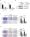
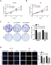
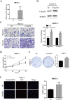
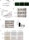
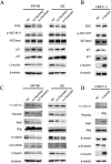
Background: Chromosome 19 open reading frame 10 (C19orf10) is a myocardial repair mediator overexpressed in hepatocellular carcinoma. However, its function and clinical value in bladder cancer (BC) have not been reported. This study aimed to investigate the role of C19orf10 in BC progression and explore underlying mechanisms. Methods: C19orf10 expression in BC tissues and human BC cell lines was assessed by reverse transcription-quantitative polymerase chain reaction (RT-qPCR) and western blot analysis. The correlation between the C19orf10 protein levels determined by immunohistochemical staining and the clinicopathological characteristics of 192 BC patients was evaluated. BC cell lines SW780, J82 and UMUC-3 were transfected with small interfering RNA (siRNA) targeting C19orf10 or plasmids overexpressing C19orf10. Cell proliferation, migration and invasion were measured by Cell Counting Kit-8, Colony formation, EdU incorporation and Transwell assays. The effect of small hairpin RNA (shRNA)-mediated stable C19orf10 knockdown on tumor formation was assessed in a xenograft mouse model. The expressions of epithelial-mesenchymal transition (EMT) markers, PI3K/AKT and Wnt/β-catenin signaling pathways-related molecules were determined by western blot assay. Results: C19orf10 was significantly upregulated in the BC tissues and a panel of human BC cell lines. High expression of C19orf10 was positively associated with malignant behaviors in BC. C19orf10 knockdown inhibited cell proliferation, migration, and invasion in SW780 and J82 cells, while C19orf10 overexpression in UMUC-3 cells resulted in opposite effects. In addition, C19orf10 silence in SW780 cells suppressed tumor growth in xenograft mice. Moreover, C19orf10 promotes the malignant behaviors and EMT of human bladder carcinoma cells via regulating the PI3K/AKT and Wnt/β-catenin pathways. Conclusion: C19orf10 is overexpressed in BC and functions as an oncogenic driver that promotes cell proliferation and metastasis, and induces EMT of BC cells via mechanisms involving activation of the PI3K/AKT and Wnt/β-catenin pathways. This study provides valuable insight on targeting C19orf10 for BC treatment.
Keywords: C19orf10; PI3K/AKT; Wnt/β-catenin; bladder cancer; migration and invasion; proliferation.
© The author(s).
Conflict of interest statement
Competing Interests: The authors have declared that no competing interest exists.
Figures


Similar articles
-
LncRNA CARLo-7 facilitates proliferation, migration, invasion, and EMT of bladder cancer cells by regulating Wnt/β-catenin and JAK2/STAT3 signaling pathways.Transl Androl Urol. 2020 Oct;9(5):2251-2261. doi: 10.21037/tau-20-1293. Transl Androl Urol. 2020. PMID: 33209690 Free PMC article.
-
CUL4B promotes bladder cancer metastasis and induces epithelial-to-mesenchymal transition by activating the Wnt/β-catenin signaling pathway.Oncotarget. 2017 Aug 24;8(44):77241-77253. doi: 10.18632/oncotarget.20455. eCollection 2017 Sep 29. Oncotarget. 2017. PMID: 29100384 Free PMC article.
-
Down-regulated MAC30 expression inhibits breast cancer cell invasion and EMT by suppressing Wnt/β-catenin and PI3K/Akt signaling pathways.Int J Clin Exp Pathol. 2019 May 1;12(5):1888-1896. eCollection 2019. Int J Clin Exp Pathol. 2019. PMID: 31934012 Free PMC article.
-
Sulforaphane and bladder cancer: a potential novel antitumor compound.Front Pharmacol. 2023 Sep 15;14:1254236. doi: 10.3389/fphar.2023.1254236. eCollection 2023. Front Pharmacol. 2023. PMID: 37781700 Free PMC article. Review.
-
Spices and culinary herbs for the prevention and treatment of breast cancer: A comprehensive review with mechanistic insights.Cancer Pathog Ther. 2024 Jul 6;3(3):197-214. doi: 10.1016/j.cpt.2024.07.003. eCollection 2025 May. Cancer Pathog Ther. 2024. PMID: 40458304 Free PMC article. Review.
Cited by
-
Etanercept ameliorates psoriasis progression through regulating high mobility group box 1 pathway.Skin Res Technol. 2023 Apr;29(4):e13329. doi: 10.1111/srt.13329. Skin Res Technol. 2023. PMID: 37113086 Free PMC article.
-
Overexpression of zinc-finger protein 677 inhibits proliferation and invasion by and induces apoptosis in clear cell renal cell carcinoma.Bioengineered. 2022 Mar;13(3):5292-5304. doi: 10.1080/21655979.2022.2038891. Bioengineered. 2022. PMID: 35164660 Free PMC article.
-
Cellular hierarchy framework based on single-cell and bulk RNA sequencing reveals fatty acid metabolic biomarker MYDGF as a therapeutic target for ccRCC.Front Immunol. 2025 Jun 5;16:1615601. doi: 10.3389/fimmu.2025.1615601. eCollection 2025. Front Immunol. 2025. PMID: 40539074 Free PMC article.
-
Myeloid-derived growth factor in diseases: structure, function and mechanisms.Mol Med. 2024 Jul 19;30(1):103. doi: 10.1186/s10020-024-00874-z. Mol Med. 2024. PMID: 39030488 Free PMC article. Review.
-
The Rapid Activation of MYDGF Is Critical for Cell Survival in the Acute Phase of Retinal Regeneration in Fish.Int J Mol Sci. 2025 Jul 27;26(15):7251. doi: 10.3390/ijms26157251. Int J Mol Sci. 2025. PMID: 40806387 Free PMC article.
References
-
- Siegel RL, Miller KD, Jemal A. Cancer statistics, 2020. CA Cancer J Clin. 2020;70(1):7–30. - PubMed
-
- Chen W, Zheng R, Baade PD, Zhang S, Zeng H, Bray F, Jemal A, Yu XQ, He J. Cancer statistics in China, 2015. CA Cancer J Clin. 2016;66(2):115–132. - PubMed
-
- Jacobs BL, Lee CT, Montie JE. Bladder cancer in 2010: how far have we come? CA Cancer J Clin. 2010;60(4):244–272. - PubMed
LinkOut - more resources
Full Text Sources

