

Intratumoral expression of interleukin 23 variants using oncolytic vaccinia virus elicit potent antitumor effects on multiple tumor models via tumor microenvironment modulation
- PMID: 34093846
- PMCID: PMC8171085
- DOI: 10.7150/thno.56494
Intratumoral expression of interleukin 23 variants using oncolytic vaccinia virus elicit potent antitumor effects on multiple tumor models via tumor microenvironment modulation
Abstract
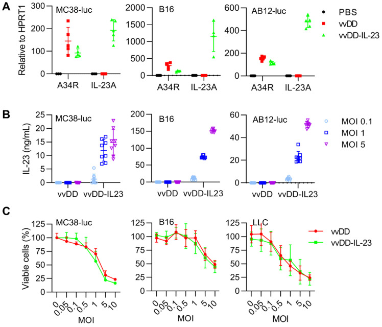
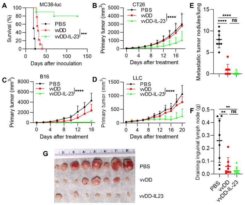
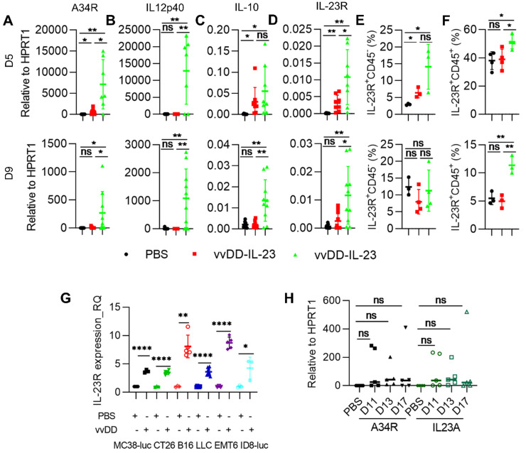
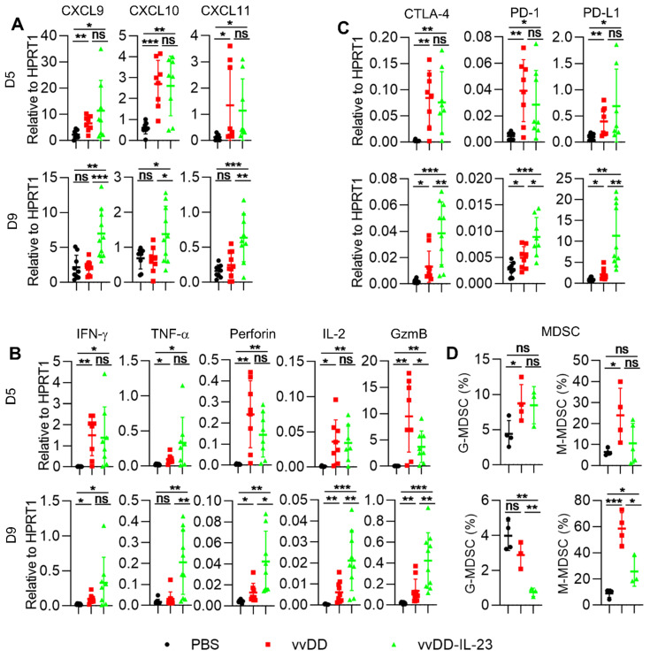
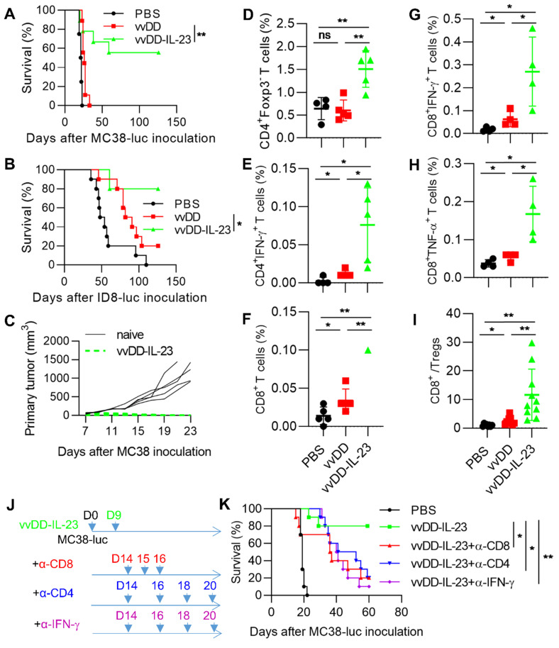
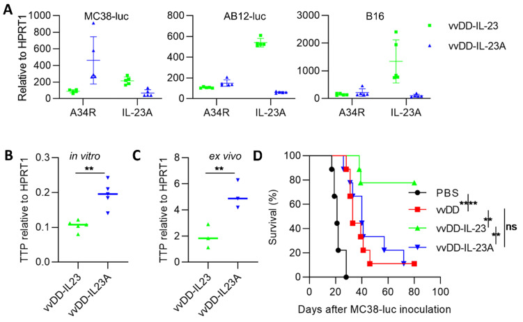
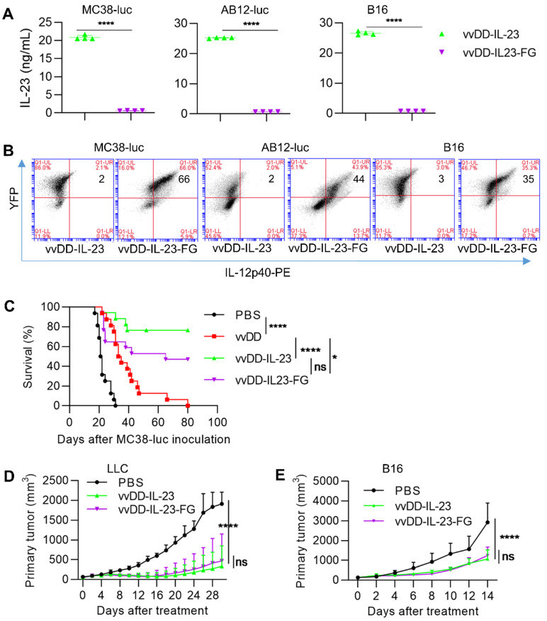
Background: Newly emerging cancer immunotherapy has led to significant progress in cancer treatment; however, its efficacy is limited in solid tumors since the majority of them are "cold" tumors. Oncolytic viruses, especially when properly armed, can directly target tumor cells and indirectly modulate the tumor microenvironment (TME), resulting in "hot" tumors. These viruses can be applied as a cancer immunotherapy approach either alone or in combination with other cancer immunotherapies. Cytokines are good candidates to arm oncolytic viruses. IL-23, an IL-12 cytokine family member, plays many roles in cancer immunity. Here, we used oncolytic vaccinia viruses to deliver IL-23 variants into the tumor bed and explored their activity in cancer treatment on multiple tumor models. Methods: Oncolytic vaccinia viruses expressing IL-23 variants were generated by homologue recombination. The characteristics of these viruses were in vitro evaluated by RT-qPCR, ELISA, flow cytometry and cytotoxicity assay. The antitumor effects of these viruses were evaluated on multiple tumor models in vivo and the mechanisms were investigated by RT-qPCR and flow cytometry. Results: IL-23 prolonged viral persistence, probably mediated by up-regulated IL-10. The sustainable IL-23 expression and viral oncolysis elevated the expression of Th1 chemokines and antitumor factors such as IFN-γ, TNF-α, Perforin, IL-2, Granzyme B and activated T cells in the TME, transforming the TME to be more conducive to antitumor immunity. This leads to a systemic antitumor effect which is dependent on CD8+ and CD4+ T cells and IFN-γ. Oncolytic vaccinia viruses could not deliver stable IL-23A to the tumor, attributed to the elevated tristetraprolin which can destabilize the IL-23A mRNA after the viral treatment; whereas vaccinia viruses could deliver membrane-bound IL-23 to elicit a potent antitumor effect which might avoid the possible toxicity normally associated with systemic cytokine exposure. Conclusion: Either secreted or membrane-bound IL-23-armed vaccinia virus can induce potent antitumor effects and IL-23 is a candidate cytokine to arm oncolytic viruses for cancer immunotherapy.
Keywords: IL-23; cancer immunotherapy; efficacy.; oncolytic virus; tumor microenvironment.
© The author(s).
Conflict of interest statement
Competing Interests: The authors have declared that no competing interest exists.
Figures
Similar articles
-
Enhanced antitumor efficacy of a novel oncolytic vaccinia virus encoding a fully monoclonal antibody against T-cell immunoglobulin and ITIM domain (TIGIT).EBioMedicine. 2021 Feb;64:103240. doi: 10.1016/j.ebiom.2021.103240. Epub 2021 Feb 10. EBioMedicine. 2021. PMID: 33581644 Free PMC article.
-
Novel oncolytic vaccinia virus armed with interleukin-27 is a potential therapeutic agent for the treatment of murine pancreatic cancer.J Immunother Cancer. 2025 May 11;13(5):e010341. doi: 10.1136/jitc-2024-010341. J Immunother Cancer. 2025. PMID: 40350204 Free PMC article.
-
Expression of CCL19 from oncolytic vaccinia enhances immunotherapeutic potential while maintaining oncolytic activity.Neoplasia. 2012 Dec;14(12):1115-21. doi: 10.1593/neo.121272. Neoplasia. 2012. PMID: 23308044 Free PMC article.
-
Preclinical and clinical trials of oncolytic vaccinia virus in cancer immunotherapy: a comprehensive review.Cancer Biol Med. 2023 Aug 23;20(9):646-61. doi: 10.20892/j.issn.2095-3941.2023.0202. Cancer Biol Med. 2023. PMID: 37615308 Free PMC article. Review.
-
Oncolytic Virus Encoding a Master Pro-Inflammatory Cytokine Interleukin 12 in Cancer Immunotherapy.Cells. 2020 Feb 10;9(2):400. doi: 10.3390/cells9020400. Cells. 2020. PMID: 32050597 Free PMC article. Review.
Cited by
-
Advances of oncolytic vaccinia viruses armed with interleukin in tumor therapy.Front Oncol. 2025 May 21;15:1594621. doi: 10.3389/fonc.2025.1594621. eCollection 2025. Front Oncol. 2025. PMID: 40469178 Free PMC article. Review.
-
Improving cancer immunotherapy by rationally combining oncolytic virus with modulators targeting key signaling pathways.Mol Cancer. 2022 Oct 12;21(1):196. doi: 10.1186/s12943-022-01664-z. Mol Cancer. 2022. PMID: 36221123 Free PMC article. Review.
-
Senecavirus A as an Oncolytic Virus: Prospects, Challenges and Development Directions.Front Oncol. 2022 Mar 17;12:839536. doi: 10.3389/fonc.2022.839536. eCollection 2022. Front Oncol. 2022. PMID: 35371972 Free PMC article. Review.
-
Intratumoural microbiota: a new frontier in cancer development and therapy.Signal Transduct Target Ther. 2024 Jan 10;9(1):15. doi: 10.1038/s41392-023-01693-0. Signal Transduct Target Ther. 2024. PMID: 38195689 Free PMC article. Review.
-
The gamble between oncolytic virus therapy and IFN.Front Immunol. 2022 Aug 25;13:971674. doi: 10.3389/fimmu.2022.971674. eCollection 2022. Front Immunol. 2022. PMID: 36090998 Free PMC article. Review.
References
-
- Groeneveldt C, van Hall T, van der Burg SH, Ten Dijke P, van Montfoort N. Immunotherapeutic Potential of TGF-beta Inhibition and Oncolytic Viruses. Trends Immunol. 2020;41:406–20. - PubMed
-
- Chen DS, Mellman I. Elements of cancer immunity and the cancer-immune set point. Nature. 2017;541:321–30. - PubMed
-
- van der Woude LL, Gorris MAJ, Halilovic A, Figdor CG, de Vries IJM. Migrating into the Tumor: a Roadmap for T Cells. Trends Cancer. 2017;3:797–808. - PubMed
Publication types
MeSH terms
Substances
Grants and funding
LinkOut - more resources
Full Text Sources
Research Materials
