

Glenoid Bone Loss in Shoulder Instability: Superiority of Three-Dimensional Computed Tomography over Two-Dimensional Magnetic Resonance Imaging Using Established Methodology
- PMID: 34094013
- PMCID: PMC8173237
- DOI: 10.4055/cios20097
Glenoid Bone Loss in Shoulder Instability: Superiority of Three-Dimensional Computed Tomography over Two-Dimensional Magnetic Resonance Imaging Using Established Methodology
Abstract
Backgroud: Recent literature suggests that three-dimensional magnetic resonance imaging (3D MRI) can replace 3D computed tomography (3D CT) when evaluating glenoid bone loss in patients with shoulder instability. We aimed to examine if 2D MRI in conjunction with a validated predictive formula for assessment of glenoid height is equivalent to the gold standard 3D CT scans for patients with recurrent glenohumeral instability.
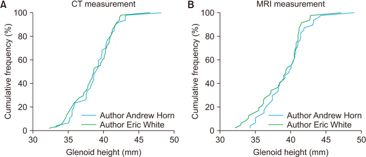
Methods: Patients with recurrent shoulder instability and available imaging were retrospectively reviewed. Glenoid height on 3D CT and 2D MRI was measured by two blinded raters. Difference and equivalence testing were performed using a paired t-test and two one-sided tests, respectively. The interclass correlation coefficient (ICC) was used to test for interrater reliability, and percent agreement between the measurements of one reviewer was used to assess intrarater reliability.
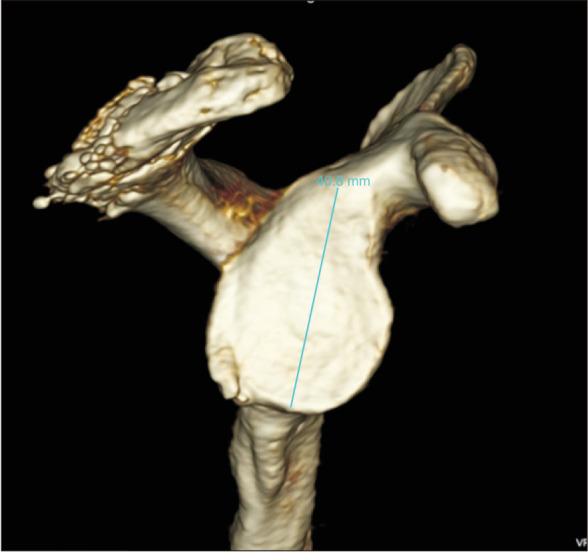
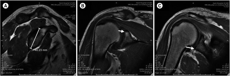
Results: Using an equivalence margin of 1 mm, 3D CT and 2D MRI were found to be different (p = 0.123). The mean glenoid height was significantly different when measured on 2D MRI (39.09 ± 2.93 mm) compared to 3D CT (38.71 ± 2.89 mm) (p = 0.032). The mean glenoid width was significantly different between 3D CT (30.13 ± 2.43 mm) and 2D MRI (27.45 ± 1.72 mm) (p < 0.001). The 3D CT measurements had better interrater agreement (ICC, 0.91) than 2D MRI measurements (ICC, 0.8). intrarater agreement was also higher on CT.
Conclusions: Measurements of glenoid height using 3D CT and 2D MRI with subsequent calculation of the glenoid width using a validated methodology were not equivalent, and 3D CT was superior. Based on the validated methods for the measurement of glenoid bone loss on advanced imaging studies, 3D CT study must be preferred over 2D MRI in order to estimate the amount of glenoid bone loss in candidates for shoulder stabilization surgery and to assist in surgical decision-making.
Keywords: Computed tomography dislocation; Glenohumeral; Images; Magnetic resonance imaging; Three dimensional imaging.
Copyright © 2021 by The Korean Orthopaedic Association.
Conflict of interest statement
CONFLICT OF INTEREST: No potential conflict of interest relevant to this article was reported.
Figures
References
-
- Stillwater L, Koenig J, Maycher B, Davidson M. 3D-MR vs. 3D-CT of the shoulder in patients with glenohumeral instability. Skeletal Radiol. 2017;46(3):325–331. - PubMed
-
- Beaulieu-Jones BR, Peebles LA, Golijanin P, et al. Characterization of posterior glenoid bone loss morphology in patients with posterior shoulder instability. Arthroscopy. 2019;35(10):2777–2784. - PubMed
-
- Boileau P, Villalba M, Hery JY, Balg F, Ahrens P, Neyton L. Risk factors for recurrence of shoulder instability after arthroscopic Bankart repair. J Bone Joint Surg Am. 2006;88(8):1755–1763. - PubMed
-
- Levy DM, Cole BJ, Bach BR., Jr History of surgical intervention of anterior shoulder instability. J Shoulder Elbow Surg. 2016;25(6):e139–e150. - PubMed
