. 2020 Aug 17;11(35):9501-9512.
doi: 10.1039/d0sc04199h.

Determination of the glycoprotein specificity of lectins on cell membranes through oxidative proteomics

Affiliations

Determination of the glycoprotein specificity of lectins on cell membranes through oxidative proteomics

Yixuan Xie et al. Chem Sci. .

Abstract

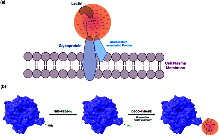
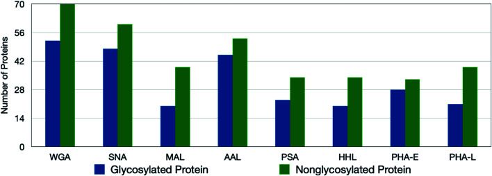
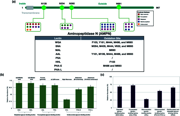
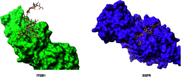
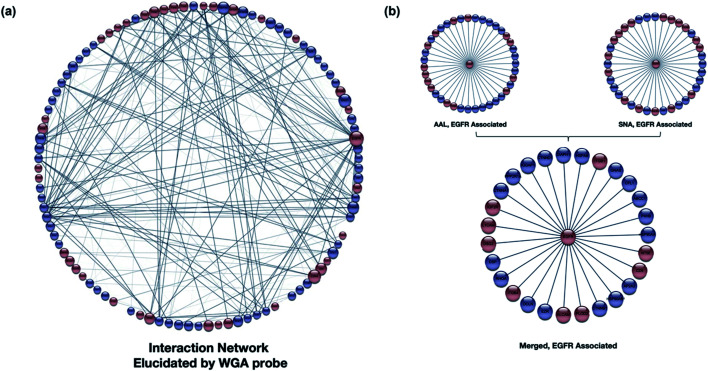
The cell membrane is composed of a network of glycoconjugates including glycoproteins and glycolipids that presents a dense matrix of carbohydrates playing critical roles in many biological processes. Lectin-based technology has been widely used to characterize glycoconjugates in tissues and cell lines. However, their specificity toward their putative glycan ligand and sensitivity in situ have been technologically difficult to study. Additionally, because they recognize primarily glycans, the underlying glycoprotein targets are generally not known. In this study, we employed lectin proximity oxidative labeling (Lectin PROXL) to identify cell surface glycoproteins that contain glycans that are recognized by lectins. Commonly used lectins were modified with a probe to produce hydroxide radicals in the proximity of the labeled lectins. The underlying polypeptides of the glycoproteins recognized by the lectins are oxidized and identified by the standard proteomic workflow. As a result, approximately 70% of identified glycoproteins were oxidized in situ by all the lectin probes, while only 5% of the total proteins were oxidized. The correlation between the glycosites and oxidation sites demonstrated the effectiveness of the lectin probes. The specificity and sensitivity of each lectin were determined using site-specific glycan information obtained through glycomic and glycoproteomic analyses. Notably, the sialic acid-binding lectins and the fucose-binding lectins had higher specificity and sensitivity compared to other lectins, while those that were specific to high mannose glycans have poor sensitivity and specificity. This method offers an unprecedented view of the interactions of lectins with specific glycoproteins as well as protein networks that are mediated by specific glycan types on cell membranes.


Conflict of interest statement

There are no conflicts with this report.

Figures

Fig. 1
Fig. 1. (a) A representation of the lectin probe attached to a target glycan and oxidizing the underlying polypeptide scaffold. The Fe(iii)-modified lectin probes were the cell supernatant and allowed to interact with the glycan target. Hydroxyl radicals were produced at the metal site creating a concentration of radicals that oxidize the proteins in proximity. The membrane proteins are isolated and analyzed by LC-MS. Oxidized proteins are identified and quantified. (b) The modification involved the introduction of the azido group into a primary amine on lysine followed by conjugation of DBCO-FeBABE to the lectin via copper-free “click” chemistry.
Fig. 2
Fig. 2. The number of oxidized glycoproteins and nonglycosylated proteins in PNT2 cell lines using lectin probes. Although more nonglycosylated proteins are oxidized, the degree of oxidation on the target glycoproteins is significantly greater.
Fig. 3
Fig. 3. (a) The relationship between the sites of glycosylation and the sites of oxidation on an example protein, AMPN. The oxidation site on the glycoprotein was highly dependent on the distribution of different types of glycans. (b) The glycoprotein specificity of lectins on PNT2 cells. The specificity of the lectins was determined as the number of oxidized glycoproteins containing the putative glycan structure versus all oxidized glycoproteins. Most of the lectins showed high specificity towards the putative target glycoproteins (>70%). The lectin HHL was the exception, which recognized only 30% high mannose glycans. The error bars were obtained based on triplicate results. (c) The sensitivity of each lectin was determined by the number of oxidized glycoproteins containing the putative target versus the total number of glycoproteins (oxidized and not oxidized) containing the putative target. The lectins were grouped because linkages of, for example, the fucose were not known. The sensitivity is highest for AAL and PSA (fucose binding lectins) and lowest for HHL (mannose binding lectins).
Fig. 4
Fig. 4. The three-dimensional structures of glycoproteins ITGB1 (integrin beta-1, PDB: 3VI3) and EGFR (epidermal growth factor receptor, PDB: 1NQL) containing the glycan Man(3)Gal(2)GlcNAc(4)Fuc(1)Sia(1). The glycoprotein models were built using Glycam (http://glycam.org). The core fucose on EGFR was predicted to be non-accessible to the PSA lectin.
Fig. 5
Fig. 5. (a) The WGA interaction network was produced using STRING (https://string-db.org). The software assigns interaction lines when known interactions (literature) are present. The glycoproteins (red) and nonglycosylated proteins (blue) are shown with their respective interactions with the size of the node proportional to the number of interactions. The weight of each protein connection showed the confidence in the interactions. (b) The interaction network of an example protein, EGFR-associated proteins as revealed by AAL and SNA. Over 80% overlap was observed in the two interaction networks illustrating the validity of the method.

Similar articles

Cited by

References

    1. Ruhaak L. R. Xu G. Li Q. Goonatilleke E. Lebrilla C. B. Mass Spectrometry Approaches to Glycomic and Glycoproteomic Analyses. Chem. Rev. 2018;118(17):7886–7930. doi: 10.1021/acs.chemrev.7b00732. - DOI - PMC - PubMed
    1. Paszek M. J. DuFort C. C. Rossier O. Bainer R. Mouw J. K. Godula K. Hudak J. E. Lakins J. N. Wijekoon A. C. Cassereau L. Rubashkin M. G. Magbanua M. J. Thorn K. S. Davidson M. W. Rugo H. S. Park J. W. Hammer D. A. Giannone G. Bertozzi C. R. Weaver V. M. The cancer glycocalyx mechanically primes integrin-mediated growth and survival. Nature. 2014;511(7509):319–325. doi: 10.1038/nature13535. - DOI - PMC - PubMed
    1. An H. J. Kronewitter S. R. de Leoz M. L. A. Lebrilla C. B. Glycomics and disease markers. Curr. Opin. Chem. Biol. 2009;13(5):601–607. doi: 10.1016/j.cbpa.2009.08.015. - DOI - PMC - PubMed
    1. Janik M. E. Lityńska A. Vereecken P. Cell migration—The role of integrin glycosylation. Biochim. Biophys. Acta, Gen. Subj. 2010;1800(6):545–555. doi: 10.1016/j.bbagen.2010.03.013. - DOI - PubMed
    1. Langer M. D. Guo H. Shashikanth N. Pierce J. M. Leckband D. E. N-glycosylation alters cadherin-mediated intercellular binding kinetics. J. Cell Sci. 2012;125(10):2478. doi: 10.1242/jcs.101147. - DOI - PubMed