. 2021 May 21:11:694957.
doi: 10.3389/fcimb.2021.694957. eCollection 2021.

Von Willebrand Factor Facilitates Intravascular Dissemination of Microsporidia Encephalitozoon hellem

Affiliations

Von Willebrand Factor Facilitates Intravascular Dissemination of Microsporidia Encephalitozoon hellem

Jialing Bao et al. Front Cell Infect Microbiol. .

Abstract

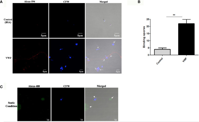
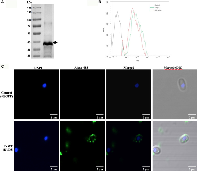
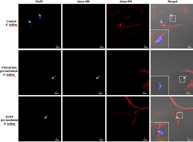
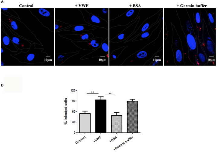
Microsporidia are a group of spore-forming, fungus-related pathogens that can infect both invertebrates and vertebrates including humans. The primary infection site is usually digestive tract, but systemic infections occur as well and cause damages to organs such as lung, brain, and liver. The systemic spread of microsporidia may be intravascular, requiring attachment and colonization in the presence of shear stress. Von Willebrand Factor (VWF) is a large multimeric intravascular protein and the key attachment sites for platelets and coagulation factors. Here in this study, we investigated the interactions between VWF and microsporidia Encephalitozoon hellem (E. hellem), and the modulating effects on E. hellem after VWF binding. Microfluidic assays showed that E. hellem binds to ultra-large VWF strings under shear stress. In vitro germination assay and infection assay proved that E. hellem significantly increased the rates of germination and infection, and these effects would be reversed by VWF blocking antibody. Mass spectrometry analysis further revealed that VWF-incubation altered various aspects of E. hellem including metabolic activity, levels of structural molecules, and protein maturation. Our findings demonstrated that VWF can bind microsporidia in circulation, and modulate its pathogenicity, including promoting germination and infection rate. VWF facilitates microsporidia intravascular spreading and systemic infection.

Keywords: Encephalitozoon hellem; infection; intravascular dissemination; microsporidia; von Willebrand factor.


Conflict of interest statement

The authors declare that the research was conducted in the absence of any commercial or financial relationships that could be construed as a potential conflict of interest.

Figures

Figure 1
Figure 1
E. hellem spores attach to FL-VWF under flow. (A) Representative images of control protein (BSA, top) or FL-VWF (bottom), both at 20 µg/ml were perfused through the microfluidic chamber with E. hellem spores (105 cells/ml) under flow at 5 dyn/cm2, respectively. The channels were then washed, fixed and stained by Alexa-594-labeled anti-VWF antibody and calcofluor-white (CFW). The fluorescent microscopy analysis showed that the VWF formed ultra large multimers under flow (red), and E. hellem spores (blue) attached to the strings of ultra large VWF strings, as pointed out by white arrows in the right figures. (Scale bar = 5 µm). (B) The number of binding spores in the channels were calculated, based on three independent studies with 8 random fields for each study (F(1,23) = 2.25, **P <0.01). (C) Under static conditions with no shear, the FL-VWF clumped and aggregated together (green). The E. hellem spores (blue) are not able to bind to clumped VWF.
Figure 2
Figure 2
VWF-D’D3 binds to E. hellem spores. (A) Coomassie staining of recombinant VWF-D’D3. Arrow shows the major protein size at the expected size ~40 kDa. (B) Flow cytometry analysis of D’D3 binding to E. hellem. E. hellem spores (1 × 10⁴ cells) were incubated respectively with, isotype antibody control (in black line), 8 ng/µl recombinant VWF-D’ D3 (in green line), and 800 ng/µl recombinant VWF-D’D3 (in red line). The result showed that with the increasing amount of recombinant VWF adding, the fluorescence signal increased as well. (C) Representative images of E. hellem spores were incubated with either control, recombinant EGFP (top) or VWF-D’D3 (bottom), both at 20 µg/ml for 30 min, then the spores were washed by PBS. After fixation, the direct interaction between VWF (green) and E. hellem (blue) was observed by fluorescent microscope (Scale bar = 2 µm).
Figure 3
Figure 3
VWF-D’D3 is key binding region for E. hellem. In microfluidic chamber, full length VWF (20 µg/ml) was perfused with shear stress of 5 dyn/cm2 for 2 min. Same concentration (105/ml) of either control (un-treated) E. hellem spores, VWF-D’D3 pre-incubated E. hellem spores, or EGFP pre-incubated E. hellem spores were then perfused through. The channels were then washed and fixed. The E. hellem spores were visualized by DAPI (blue), and the pre-incubated VWF-D’D3 which has attached to E. hellem spores were visualized by anti-His antibody followed by Alexa 488-labeled secondary antibody (green). The VWF oligomers were visualized by anti-VWF antibody followed by Alexa 594-labeled secondary antibody (red). As shown by this immunofluorescence assay, untreated E. hellem spores or un-related EGFP treated E. hellem spores were both able to attach to the VWF oligomer strings (arrows, and also shown in enlarged views in upper and bottom rows). While VWF-D’D3 pre-incubation occupy the binding site of E. hellem, thus the spores could not bind with VWF strings (arrow, and also shown in enlarged view in middle row).
Figure 4
Figure 4
VWF binding promotes E. hellem germination. (A) Representative images of E. hellem germination affected by VWF. The control group E. hellem spores were untreated by any protien; The VWF group E. hellem spores were incubated with FL-VWF; The VWF + VWF-antibody group E. hellem spores were treated by VWF together with anti-VWF antibody; The VWF + isotype antibody group E. hellem spores were treated by VWF together with isotype antibody control. All the spores from each group were then stimulated with germination buffer to further trigger germination. The E. hellem spores were then stained by DAPI, and un-germinated spores will show blue color (pointed out by arrows). (Scale bar = 5 µm). (B) Germination rates were calculated by the ratio of germinated spores over all spores, based on three independent studies with 10 random fields per study. The results showed that VWF treatment significantly promoted E. hellem spores’ germination (F(1,29) = 1.89, *P <0.05), and this effect was inhibited by VWF specific antibody (F(1, 29) = 2.09, *P <0.05).
Figure 5
Figure 5
VWF promotes E. hellem infection. (A) Representative fields of HFF cells exposed to E. hellem spores that had undergone no treatment (control) or binding of either FL-VWF or BSA or exposed to germination buffer. The spores were then added to HFF cells and culture for 12 h. The HFF cells outlines were depicted as ‘dots’ by Adobe IIIustrator CS6to the DIC images of the cells. HFF cell nuclei were stained by DAPI (blue), while the infected E. hellem was represented by FISH probe (red) (Scale bar = 10 µm). (B) Infectivity rate was the ratio of infected HFF cells over all cells, based on three independent studies with 20 random fields per study. The result showed that VWF treatment significantly increased the infection rate of E. hellem to host cells (F(1,59) = 2.42,**=P <0.01); while the un-related protein treatment of E. hellem spores had no effect on the infection ability (F(1,59) = 1.92, **P <0.01).
Figure 6
Figure 6
GO annotation and enrichment analysis of differentially expressed proteins in E. hellem post-exposure to VWF. The primary Y axis denotes the number of annotated proteins categorized to each GO term. The secondary Y axis represents the percentage of annotated proteins to each GO term in all differential proteins. GO terms are classified into three subcategories, including biological process (BP), molecular function (MF) and cellular compartment (CC). The color gradient represents the p-value; the closer to red, the smaller the p-value. The enriched proteins are categorized and showed on X axis as: 1—Protein maturation by iron-sulfur cluster transfer; 2—Protein maturation by [4Fe–4S] cluster transfer; 3—Histone deubiquitination; 4—Structural molecule activity; 5—Oxidoreductase activity; 6—Oxidoreductase activity, acting on CH2 groups; 7—Ribonucleoside-diphosphate reductase activity; 8—Trehalase activity; 9—Thiol-dependent ubiquitin-specific protease activity; 10—Glucose-6-phosphate isomerise activity; 11—Damage DNA binding; 12—Single-stranded DNA binding; 13—Thioredoxin activity; 14—Alpha trehalase activity; 15—Structural constituent of ribosome; 16—Vacuolar proton-transporting V-type ATPase.

Similar articles

References

    1. Anderson N. W., Muehlenbachs A., Arif S., Bruminhent J., Deziel P. J., Razonable R. R., et al. . (2019). A Fatal Case of Disseminated Microsporidiosis Due to Anncaliia Algerae in a Renal and Pancreas Allograft Recipient. Open Forum Infect. Dis. 6, ofz285. 10.1093/ofid/ofz285 - DOI - PMC - PubMed
    1. Barber B. E., William T., Grigg M. J., Parameswaran U., Piera K. A., Price R. N., et al. . (2015). Parasite Biomass-Related Inflammation, Endothelial Activation, Microvascular Dysfunction and Disease Severity in Vivax Malaria. PloS Pathog. 11, e1004558. 10.1371/journal.ppat.1004558 - DOI - PMC - PubMed
    1. Bukreyeva I., Angoulvant A., Bendib I., Gagnard J. C., Bourhis J. H., Dargere S., et al. . (2017). Enterocytozoon Bieneusi Microsporidiosis in Stem Cell Transplant Recipients Treated With Fumagillin. Emerging Infect. Dis. 23, 1039–1041. 10.3201/eid2306.161825 - DOI - PMC - PubMed
    1. Delgado Betancourt E., Hamid B., Fabian B. T., Klotz C., Hartmann S., Seeber F. (2019). From Entry to Early Dissemination-Toxoplasma Gondii’s Initial Encounter With Its Host. Front. Cell Infect. Microbiol. 9, 46. 10.3389/fcimb.2019.00046 - DOI - PMC - PubMed
    1. Didier E. S., Maddry J. A., Brindley P. J., Stovall M. E., Didier P. J. (2005). Therapeutic Strategies for Human Microsporidia Infections. Expert Rev. Anti-infective Ther. 3, 419–434. 10.1586/14787210.3.3.419 - DOI - PubMed

Publication types

Substances

Supplementary concepts