. 2021 May 20:9:654163.
doi: 10.3389/fcell.2021.654163. eCollection 2021.

Comparative Genomics of Peroxisome Biogenesis Proteins: Making Sense of the PEX Proteins

Affiliations

Comparative Genomics of Peroxisome Biogenesis Proteins: Making Sense of the PEX Proteins

Renate L M Jansen et al. Front Cell Dev Biol. .

Abstract

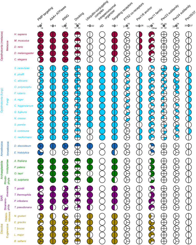
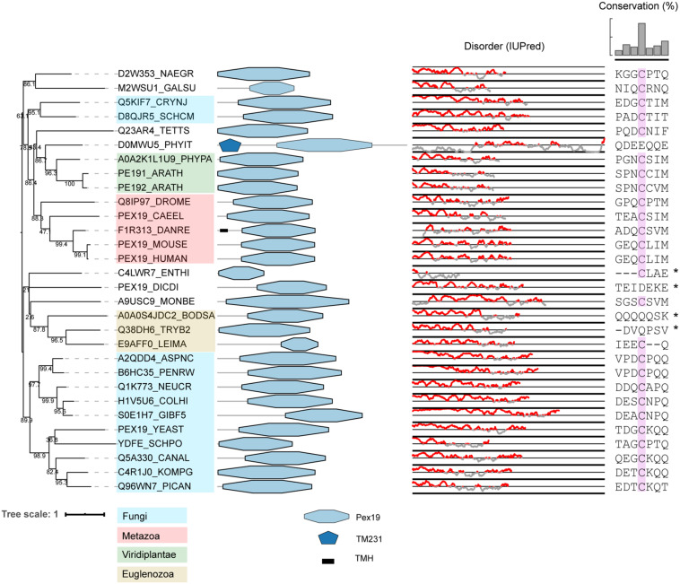
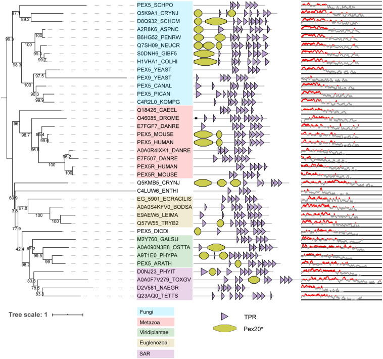
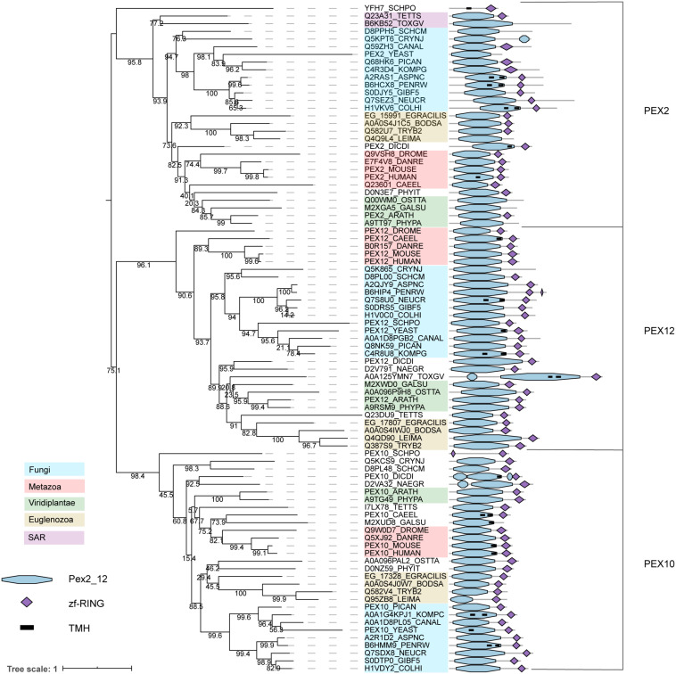
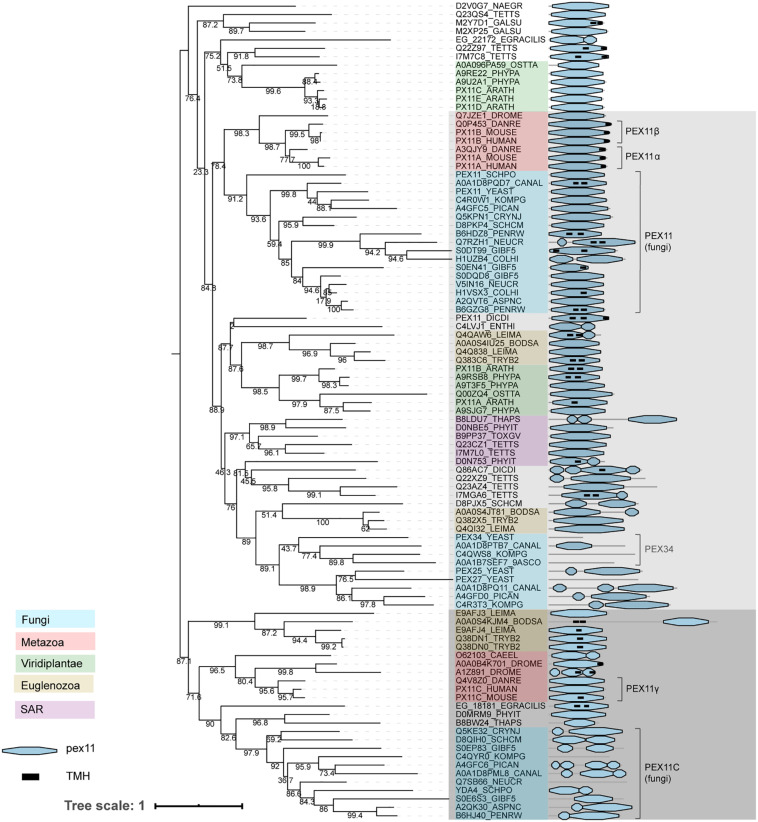
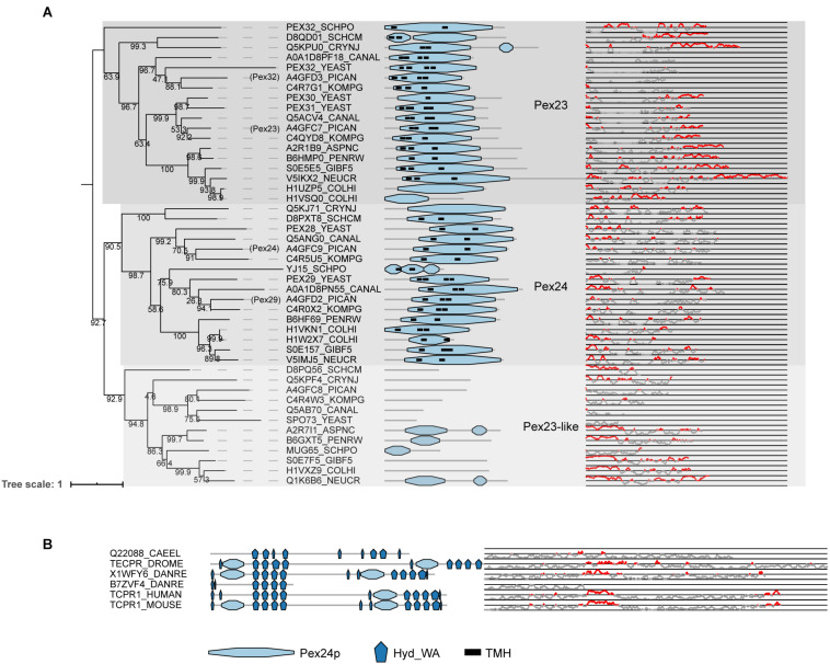
PEX genes encode proteins involved in peroxisome biogenesis and proliferation. Using a comparative genomics approach, we clarify the evolutionary relationships between the 37 known PEX proteins in a representative set of eukaryotes, including all common model organisms, pathogenic unicellular eukaryotes and human. A large number of previously unknown PEX orthologs were identified. We analyzed all PEX proteins, their conservation and domain architecture and defined the core set of PEX proteins that is required to make a peroxisome. The molecular processes in peroxisome biogenesis in different organisms were put into context, showing that peroxisomes are not static organelles in eukaryotic evolution. Organisms that lack peroxisomes still contain a few PEX proteins, which probably play a role in alternative processes. Finally, the relationships between PEX proteins of two large families, the Pex11 and Pex23 families, were analyzed, thereby contributing to the understanding of their complicated and sometimes incorrect nomenclature. We provide an exhaustive overview of this important eukaryotic organelle.

Keywords: PEX; comparative genomics; evolution; peroxisome; protein domains.


Conflict of interest statement

The authors declare that the research was conducted in the absence of any commercial or financial relationships that could be construed as a potential conflict of interest.
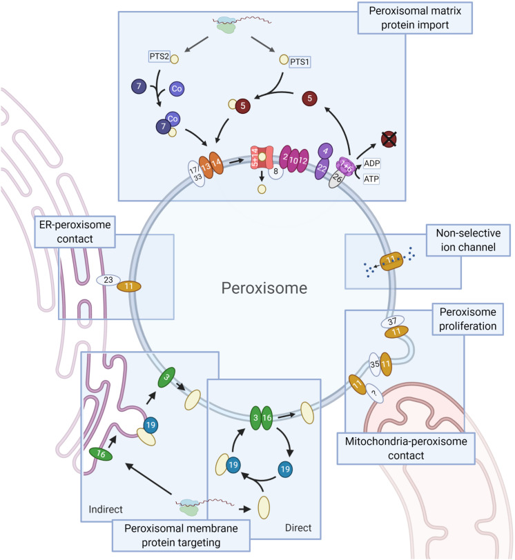
Figures

FIGURE 1
FIGURE 1
Schematic representation of the PEX proteins. Core conserved PEX proteins (shapes in dark colors, names in white), fungi-specific proteins (light, names in black) and the moderately conserved PEX protein PEX26 (gray, name in black, which is only present in Metazoa and Fungi) are depicted. Membrane proteins are ovals, soluble proteins round. Matrix protein import. Peroxisomal matrix proteins contain a peroxisomal targeting signal (PTS) that is recognized by cytosolic receptors: a C-terminal PTS1 or (less commonly) an N-terminal PTS2, recognized by PEX5 and PEX7, respectively. PTS2 import involves a co-receptor (Co): PEX5 (animals, plants, and protists), PEX18/21 (S. cerevisiae) or PEX20 (Fungi). Next, the receptor-cargo complex associates with the docking complex, consisting of PEX13/14 (and in Fungi PEX17 or PEX33). Upon cargo translocation and release, the PTS (co-)receptor is ubiquitinated and recycled. Ubiquitination involves the ubiquitin conjugating enzyme (E2) PEX4 (recruited to the membrane by PEX22) and the ubiquitin ligase (E3) activities of the RING finger complex, consisting of PEX2/10/12. Receptor extraction requires the AAA+ ATPase complex PEX1/6, which is recruited to the membrane via PEX26 (PEX15 in S. cerevisiae, APEM9 in plants – only PEX26 shown). PEX8 links the docking and RING finger complexes, and functions in receptor-cargo dissociation. Peroxisomal membrane protein (PMP) targeting involves PEX3, PEX19 and PEX16. PMPs can sort directly to peroxisomes or indirectly via the ER. In the direct pathway PEX19 acts as receptor/chaperone, while it functions at the ER in PMP sorting via the indirect pathway. The Pex11 protein family (all show as PEX11) and the fungal peroxins PEX35 and PEX37 have been mainly implicated in peroxisome proliferation. Pex11 family proteins are also present in mitochondria-peroxisome contact sites and PEX11 functions as non-selective ion channel. Members of the fungal Pex23 protein family localize to the ER and are involved in the formation of peroxisome-ER membrane contact sites. Created with BioRender.com.
FIGURE 2
FIGURE 2
Coulson plot demonstrating the presence (filled) or absence (empty) of PEX protein orthologs in 32 eukaryotic proteomes. PEX proteins are divided into functional groups (columns) including homologous and non-homologous proteins, represented by a pie. Every wedge represents a PEX protein, with the exception of the Pex11 family, where each wedge represents a main group. The PEX11 group contains among others fungal PEX11/25/27/34/36 and mammalian PEX11α/β. The PEX11C group includes fungal PEX11C and mammalian PEX11γ. Pex11 family proteins that do not belong to the either of these groups are placed in “Other.” Organisms are grouped by eukaryotic supergroup (color-coded for clarity) and kingdom. PEX proteins are designated by their number.
FIGURE 3
FIGURE 3
Phylogeny and protein features of PEX19 orthologs. The phylogeny is rooted at mid-point to ease the visualization and labels of the main taxonomic groups are colored according to the legend. Note that the topology does not necessarily reflect the actual evolutionary trajectory of such proteins. Protein domain architecture is defined by pfam annotations and transmembrane helices (TMH) according to TMHMM software. The line-dot plot, indicates the regions predicted to be disordered (red) and not disordered (gray). The sequence alignment shows the conservation of the CaaX box in PEX19 orthologs of distant eukaryotes, with ‘C’ denoting Cys, ‘a’ an aliphatic residue and ‘X’ usually being a Ser, Thr, Gln, Ala or Met. Asterisk indicates forced alignments manually.
FIGURE 4
FIGURE 4
Phylogeny and protein features of PEX5 orthologs. The phylogeny is rooted at mid-point to ease the visualization and labels of the main taxonomic groups are colored accordingly to the legend. Note that the topology does not necessarily reflect the actual evolutionary trajectory of such proteins. Protein domain architecture is defined by pfam annotations. The Pex20 is a manually generated hidden Markov model (CSM, this study). The line-dot plot indicates the regions predicted be disordered (red) and not disordered (gray).
FIGURE 5
FIGURE 5
Phylogeny and protein features of PEX2/10/12 orthologs. The phylogeny is rooted at mid-point to ease the visualization and labels of the main taxonomic groups are colored accordingly to the legend. Note that the topology does not necessarily reflect the actual evolutionary trajectory of such proteins. Protein domain architecture is defined by pfam annotations and transmembrane helix according to TMHMM software.
FIGURE 6
FIGURE 6
Phylogeny and protein features of PEX11 family proteins. The phylogeny is rooted at mid-point to ease the visualization and labels of the main taxonomic groups are colored accordingly to the legend. Note that the topology does not necessarily reflect the actual evolutionary trajectory of such proteins. Protein domain architecture is defined by pfam annotations and transmembrane helix prediction (black box). The two main groups, shaded light and dark gray, are distinguished according to the most supported and basal bootstraps and their taxonomic compositions.
FIGURE 7
FIGURE 7
Phylogeny and protein features of Pex23 protein family and TECPR1 proteins. (A) Phylogeny and protein features of the fungal Pex23 protein family. The phylogeny is rooted at mid-point to ease the visualization. The main phylogenetic groups are named and highlighted according the protein names of O. polymorpha, indicated between brackets. Protein domain architecture is defined by pfam annotations and transmembrane helix according to TMHMM software. The Pex24p pfam domain contains the DysF motifs. The line-dot plot, indicates the region predicted be disordered (red) and not disordered (gray). (B) Protein features of TECPR1 protein family from Metazoa.

References

    1. Agne B., Meindl N. M., Niederhoff K., Einwächter H., Rehling P., Sickmann A., et al. (2003). Pex8p: an intraperoxisomal organizer of the peroxisomal import machinery. Mol. Cell 11 635–646. 10.1016/s1097-2765(03)00062-5 - DOI - PubMed
    1. Agrawal G., Fassas S. N., Xia Z., Subramani S. (2016). Distinct requirements for intra-ER sorting and budding of peroxisomal membrane proteins from the ER. J. Cell Biol. 212 335–348. 10.1083/jcb.201506141 - DOI - PMC - PubMed
    1. Amery L., Sano H., Mannaerts G. P., Snider J., van Looy J., Fransen M., et al. (2001). Identification of PEX5p-related novel peroxisome-targeting signal 1 (PTS1)-binding proteins in mammals. Biochem. J. 357 635–646. 10.1042/bj3570635 - DOI - PMC - PubMed
    1. Ban N., Beckmann R., Cate J. H., Dinman J. D., Dragon F., Ellis S. R., et al. (2014). A new system for naming ribosomal proteins. Curr. Opin. Struct. Biol. 24 165–169. - PMC - PubMed
    1. Barros-Barbosa A., Ferreira M. J., Rodrigues T. A., Pedrosa A. G., Grou C. P., Pinto M. P., et al. (2019a). Membrane topologies of PEX 13 and PEX 14 provide new insights on the mechanism of protein import into peroxisomes. FEBS J. 286 205–222. 10.1111/febs.14697 - DOI - PubMed